You have no items in your shopping cart.
Cart summary
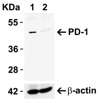
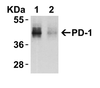
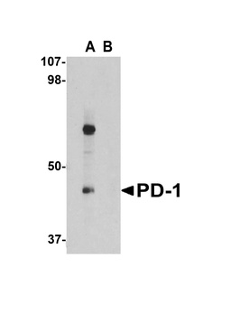
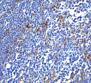
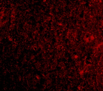
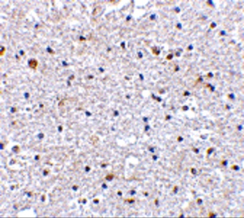
PD1 antibody
Catalog Number: orb758881
| Catalog Number | orb758881 |
|---|---|
| Category | Antibodies |
| Description | Rabbit monoclonal antibody to PD1 |
| Species/Host | Hamster |
| Clonality | Monoclonal |
| Clone Number | J43 |
| Tested applications | ELISA, FC, IHC, IP |
| Reactivity | Mouse |
| Isotype | IgG |
| Immunogen | This antibody was raised by immunising Armenian hamsters with B12 cells, a PD-1 cDNA transfectant of BHK cells. |
| Concentration | 1 mg/ml |
| Purity | Purified |
| Conjugation | Unconjugated |
| Target | PD-1 |
| UniProt ID | Q02242 |
| Storage | Store at 4°C for up to 3 months. For longer storage, aliquot and store at -20°C. |
| Buffer/Preservatives | PBS with 0.02% Proclin 300. |
| Alternative names | CD279; PD1; PD 1; Programmed cell death protein 1; Read more... |
| Note | For research use only |
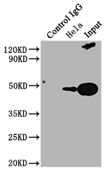
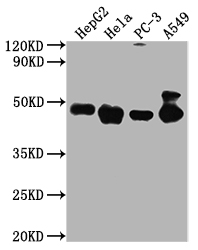
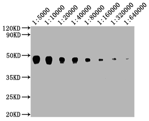
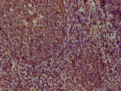
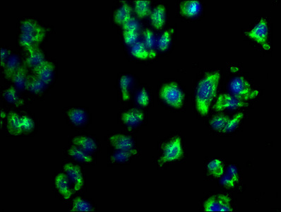
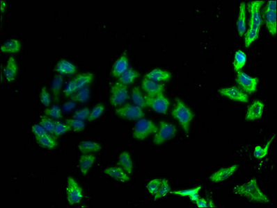
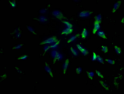
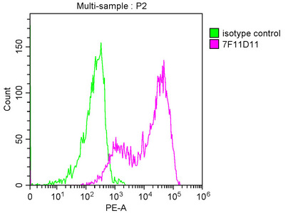
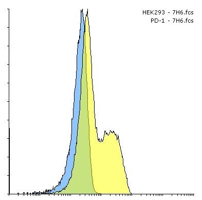
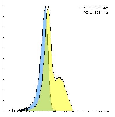
| Application notes | The specificity of this antibody has been confirmed in ELISA analysis, using PD-1 extracellular domain fusion proteins (Agata et al, 1996). Additionally, in flow cytometric analysis, this antibody reacts with PD-1 cDNA-transfected BHK and CHO cells, but not with parental BHK and CHO cells, as well as reacting with lymphocytes from PD-1 cDNA transgenic mice (Agata et al, 1996). This antibody has been used to immunoprecipitate PD-1 from lysates of PD-1 cDNA-transfected BHK and CHO cells (Agata et al, 1996), in flow cytometric quantification of CD4+PD-1+ T cells in murine spleens (Kasagi et al, 2010), and in immunohistochemical analysis of acetone-fixed murine spinal cord and brain tissue sections (Salama et al, 2003). This antibody displays diverse effects in different mouse models of disease. When administered to NZB/W F1 mice, a model of lupus-like nephritis, this antibody has been shown to delay the onset of nephritis and prolong survival, through the depletion of PD-1+ T cells (Kasagi et al, 2010). Antibody-treated NZB/W F1 mice displayed decreased numbers of PD-1+ T cells, and this antibody was confirmed to trigger complement-dependent cytotoxicity in PD-1+ T cells in vitro (Kasagi et al, 2010). Conversely, administration to experimental allergic encephalitis (EAE) and NOD diabetes mice exacerbated disease, through its neutralising activity (Salama et al, 2003; Ansari et al, 2003); this antibody has been shown in vitro to inhibit binding of both PD-L1-Ig and PD-L2-Ig to PD-1 transfected BHK cells (Ansari et al, 2003). |
| Expiration Date | 12 months from date of receipt. |
- Item 1 of 9
- Item 1 of 8
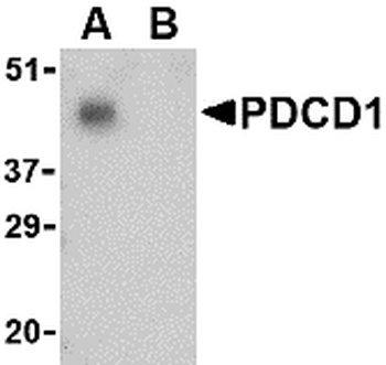
PDCD1 Antibody [orb1239732]
ELISA, IF, IHC-P, WB
Human, Mouse, Rat
Rabbit
Polyclonal
Unconjugated
0.1 mg, 0.02 mg - Item 1 of 8
- Item 1 of 7
- Item 1 of 6


Submit a review
Filter by Rating

- 5 stars

- 4 stars
- 3 stars
- 2 stars
- 1 stars