You have no items in your shopping cart.
Cart summary
Item 1 of 6
Item 1 of 6
NEK2 Antibody
Catalog Number: orb1262771

| Catalog Number | orb1262771 |
|---|---|
| Category | Antibodies |
| Description | NEK2 Antibody |
| Target | NEK2 |
| Clonality | Polyclonal |
| Isotype | Rabbit Ig |
| Conjugation | Unconjugated |
| Reactivity | Human |
| Form/Appearance | Liquid |
| Concentration | batch dependent |
| Buffer/Preservatives | Supplied in PBS with 0.09% (W/V) sodium azide. |
| Purification | This antibody is prepared by Saturated Ammonium Sulfate (SAS) precipitation followed by dialysis |
| Immunogen | This NEK2 antibody is generated from rabbits immunized with a KLH conjugated synthetic peptide between 396-426 amino acids from the Central region of human NEK2. |
| UniProt ID | P51955 |
| MW | 52 kDa |
| Tested applications | IF, IHC-P, WB |
| Application notes | For IHC-P starting dilution is: 1:100For IF starting dilution is: 1:25For WB starting dilution is: 1:1000 |
| Antibody Type | Primary Antibody |
| Storage | Maintain refrigerated at 2-8°C for up to 2 weeks. For long term storage store at -20°C in small aliquots to prevent freeze-thaw cycles. |
| Alternative names | Serine/threonine-protein kinase Nek2, HSPK 21, Nev Read more... |
| Note | For research use only |
| NCBI | P51955 |
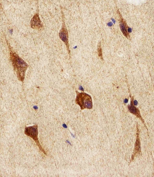
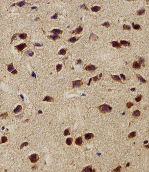
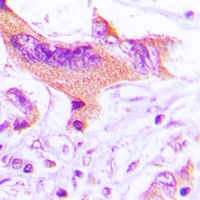
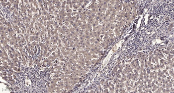
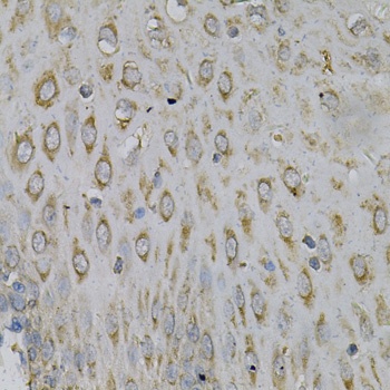
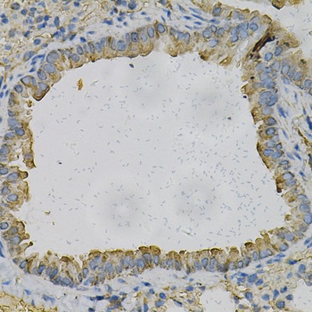
Immunohistochemical analysis of paraffin-embedded H. brain section using NEK2 Antibody. Antibody was diluted at 1:100 dilution. A peroxidase-conjugated goat anti-rabbit IgG at 1:400 dilution was used as the secondary antibody, followed by DAB staining.
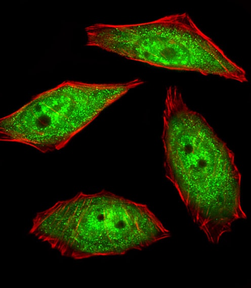
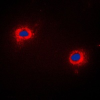
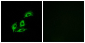
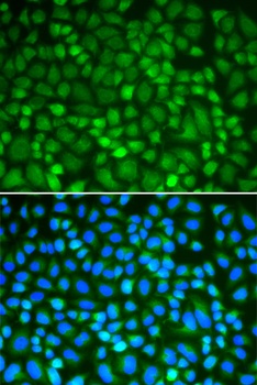
Fluorescent image of U251 cells stained with hNEK2-C410. Antibody was diluted at 1:25 dilution. An Alexa Fluor 488-conjugated goat anti-rabbit lgG at 1:400 dilution was used as the secondary antibody (green). Cytoplasmic actin was counterstained with Alexa Fluor 555 conjugated with Phalloidin (red).
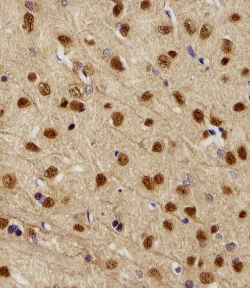
Immunohistochemical analysis of paraffin-embedded R. brain section using NEK2 Antibody. Antibody was diluted at 1:100 dilution. A peroxidase-conjugated goat anti-rabbit IgG at 1:400 dilution was used as the secondary antibody, followed by DAB staining.

Immunohistochemical analysis of paraffin-embedded M. brain section using NEK2 Antibody. Antibody was diluted at 1:100 dilution. A peroxidase-conjugated goat anti-rabbit IgG at 1:400 dilution was used as the secondary antibody, followed by DAB staining.
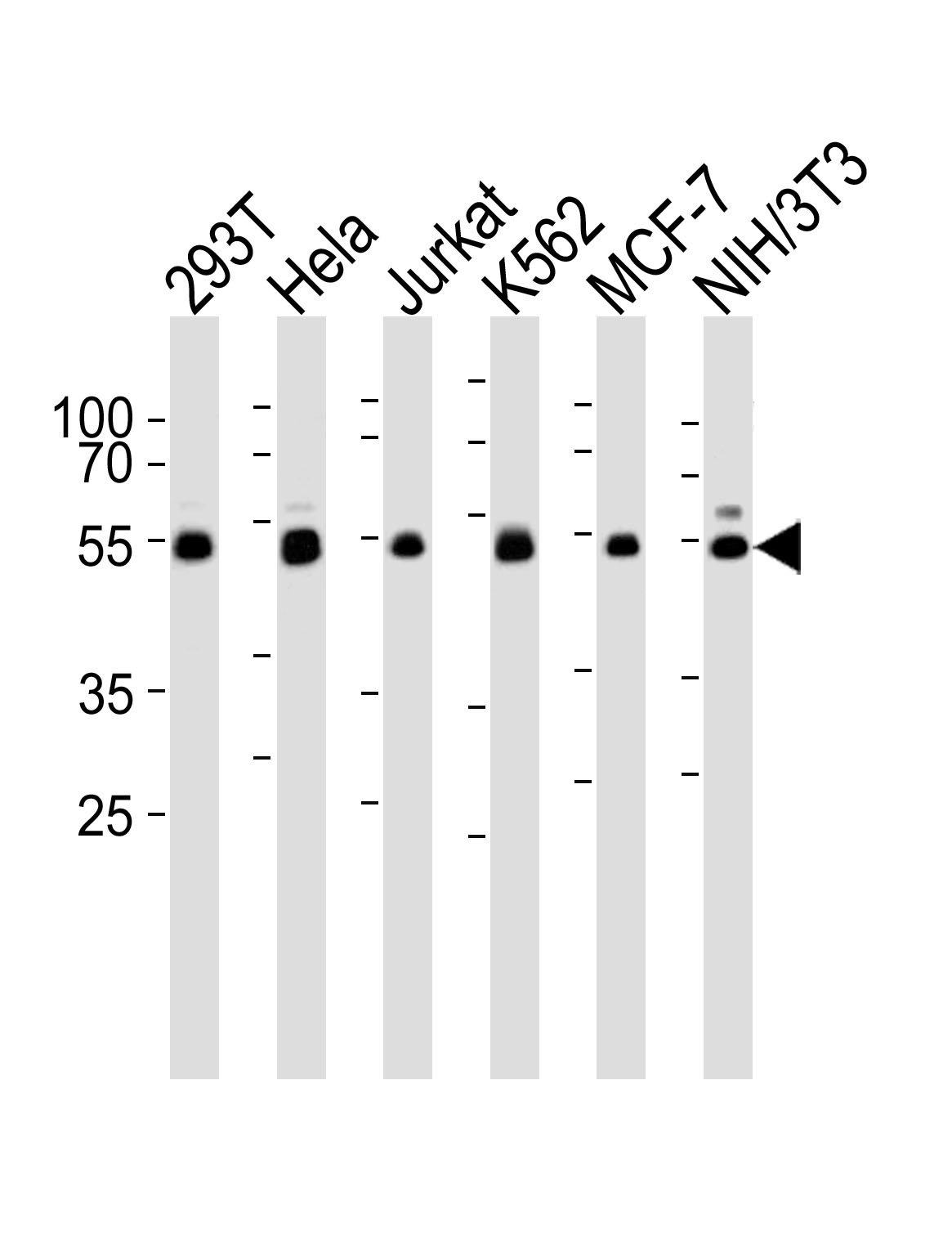
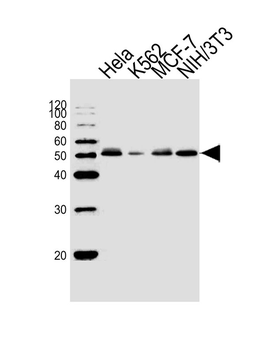
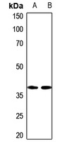
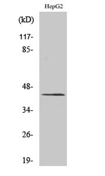
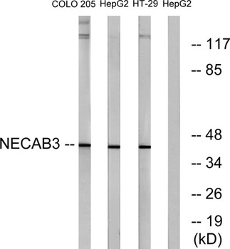
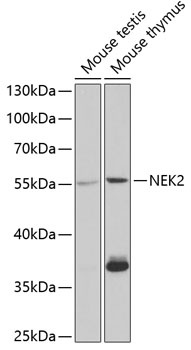
Western blot analysis of lysates from 293T, Hela, Jurkat, K562, MCF-7, and mouse NIH/3T3 cell line (from left to right), using NEK2 Antibody (C410) at 1:1000 at each lane.
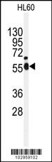
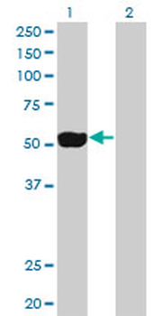
Western blot analysis of anti-NEK2 Antibody in HL60 cell line lysates (35 ug/lane)
- Item 1 of 5
- Item 1 of 3
- Item 1 of 3
Anti-NIP1 Antibody [orb215256]
IF, IH, WB
Human, Mouse, Rat
Rabbit
Polyclonal
Unconjugated
30 μl, 100 μl, 200 μl - Item 1 of 4
- Item 1 of 4