You have no items in your shopping cart.
Cart summary
Item 1 of 7
Item 1 of 7
FGFR2 Antibody
Catalog Number: orb1263054

| Catalog Number | orb1263054 |
|---|---|
| Category | Antibodies |
| Description | FGFR2 Antibody |
| Target | FGFR2 |
| Clonality | Polyclonal |
| Isotype | Rabbit IgG |
| Conjugation | Unconjugated |
| Reactivity | Human, Mouse |
| Form/Appearance | Liquid |
| Concentration | batch dependent |
| Buffer/Preservatives | Supplied in PBS with 0.09% (W/V) sodium azide. |
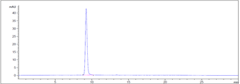
| Purification | This antibody is purified through a protein G column, followed by dialysis against PBS. |
| Immunogen | This FGFR2 antibody is generated from rabbits immunized with a KLH conjugated synthetic peptide between 22-51 amino acids from the N-terminal region of human FGFR2. |
| UniProt ID | P21802 |
| MW | 92 kDa |
| Tested applications | FC, IF, IHC-P, WB |
| Application notes | For WB starting dilution is: 1:1000-1:2000For IF starting dilution is: 1:25For IHC-P starting dilution is: 1:25For FACS starting dilution is: 1:10~50 |
| Antibody Type | Primary Antibody |
| Storage | Maintain refrigerated at 2-8°C for up to 2 weeks. For long term storage store at -20°C in small aliquots to prevent freeze-thaw cycles. |
| Alternative names | Fibroblast growth factor receptor 2, FGFR-2, K-sam Read more... |
| Note | For research use only |
| NCBI | P21802 |
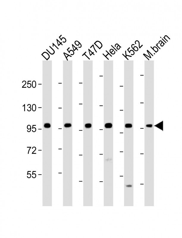
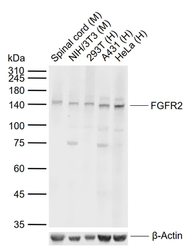
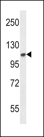
Western Blot at 1:1000-1:2000 dilution Lane 1: DU145 whole cell lysate Lane 2: A549 whole cell lysate Lane 3: T47D whole cell lysate Lane 4: Hela whole cell lysate Lane 5: K562 whole cell lysate Lane 6: M.brain whole lysate Lysates/proteins at 20 ug per lane.
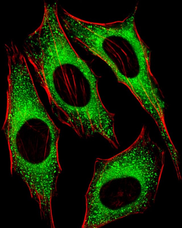
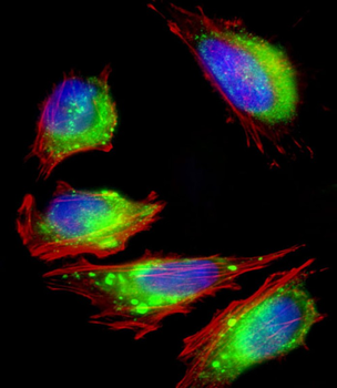
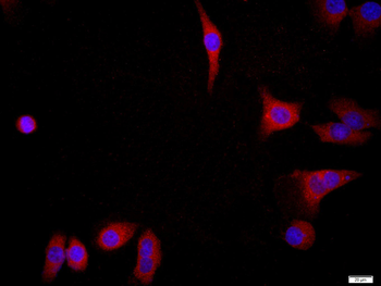
Fluorescent image of Hela cells stained with FGFR2 Antibody (N-term). Antibody was diluted at 1:25 dilution. An Alexa Fluor 488-conjugated goat anti-rabbit lgG at 1:400 dilution was used as the secondary antibody (green). Cytoplasmic actin was counterstained with Alexa Fluor 555 conjugated with Phalloidin (red).
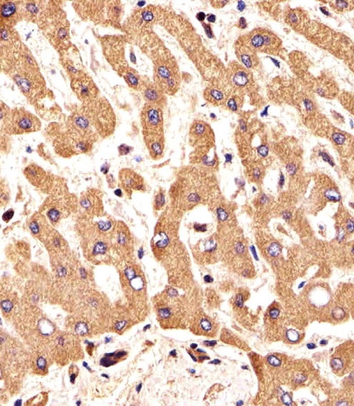
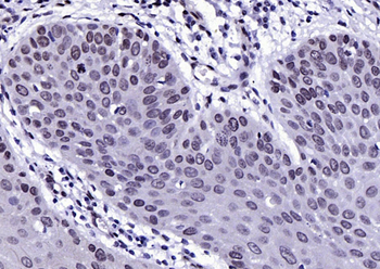
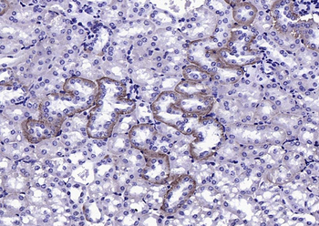
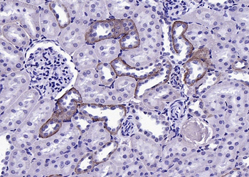
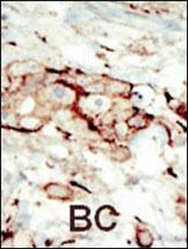
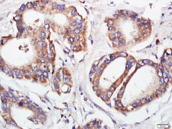
Immunohistochemical analysis of paraffin-embedded H. liver section using FGFR2 Antibody (N-term). Antibody was diluted at 1:25 dilution. A undiluted biotinylated goat polyvalent antibody was used as the secondary, followed by DAB staining.
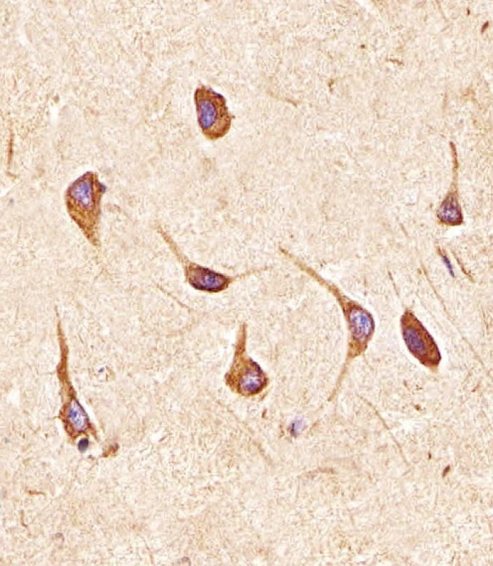
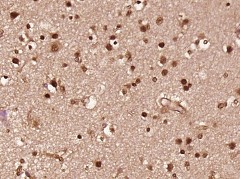
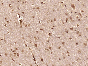
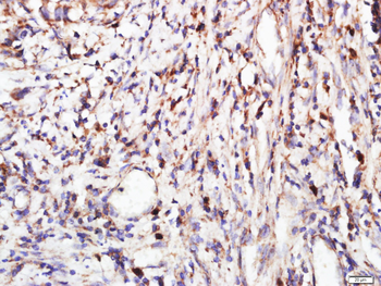
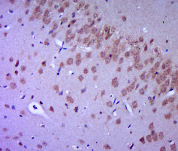
Immunohistochemical analysis of paraffin-embedded H. brain section using FGFR2 Antibody (N-term). Antibody was diluted at 1:25 dilution. A undiluted biotinylated goat polyvalent antibody was used as the secondary, followed by DAB staining.
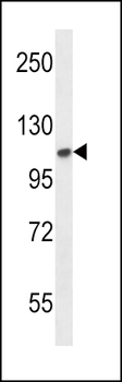
Western blot analysis in mouse NIH-3T3 cell line lysates (35 ug/lane).
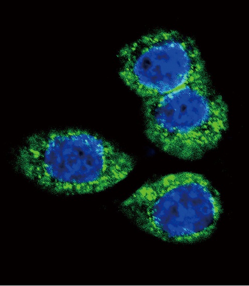
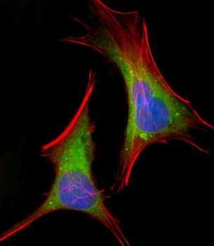
Confocal immunofluorescent analysis of FGFR2 Antibody with Hela cell followed by Alexa Fluor 488-conjugated goat anti-rabbit lgG (green).DAPI was used to stain the cell nuclear (blue).
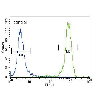
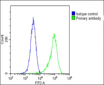
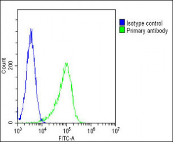
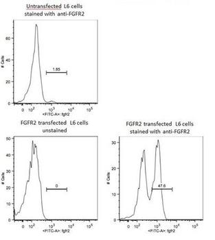
Flow cytometric analysis of NCI-H460 cells (right histogram) compared to a negative control cell (left histogram). FITC-conjugated goat-anti-rabbit secondary antibodies were used for the analysis.
- Item 1 of 7
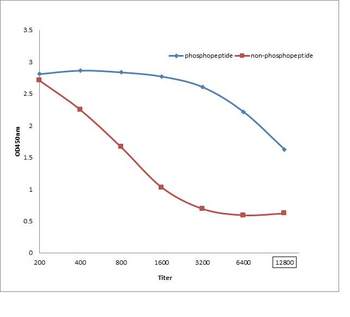
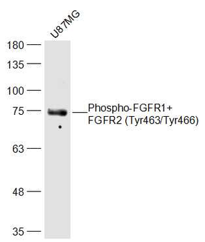
Phospho-FGFR1+FGFR2 (Tyr463/Tyr466) Rabbit Polyclonal Antibody [orb5241]
ELISA, IF, IHC-Fr, IHC-P, WB
Bovine, Equine, Gallus, Porcine, Rabbit
Human, Mouse, Rat
Rabbit
Polyclonal
Unconjugated
200 μl, 100 μl, 50 μl - Item 1 of 7
FGFR2 Antibody (N-term) [orb1929046]
FC, IF, IHC-P, WB
Human, Mouse
Rabbit
Polyclonal
Unconjugated
100 μl, 50 μl - Item 1 of 6
FGFR2 Rabbit Polyclonal Antibody [orb10656]
FC, ICC, IF, IHC-Fr, IHC-P, WB
Rat
Human, Mouse
Rabbit
Polyclonal
Unconjugated
100 μl, 200 μl, 50 μl - Item 1 of 6
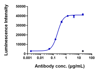
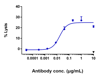
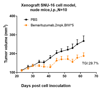
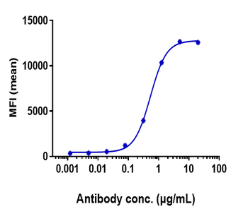
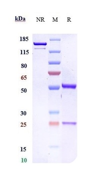
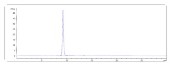
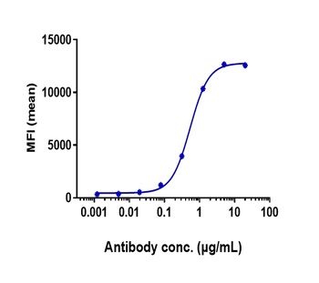
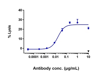
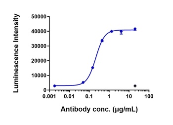
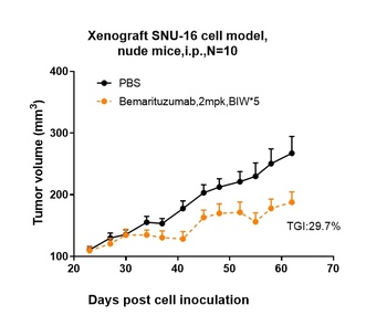
Anti-FGFR2 / CD332 Reference Antibody (bemarituzumab) [orb1817790]
ELISA, FC
Human, Monkey, Mouse
Monoclonal
Unconjugated
100 μg - Item 1 of 6
Anti-FGFR2 / CD332 Reference Antibody (bemarituzumab) [orb1806350]
ELISA, FA, FACS, Kinetics
Human
Monoclonal
Unconjugated
50 μg, 100 μg, 1 mg, 5 mg