You have no items in your shopping cart.
Cart summary
Item 1 of 6
Item 1 of 6
EphA2 Antibody
Catalog Number: orb1263100

| Catalog Number | orb1263100 |
|---|---|
| Category | Antibodies |
| Description | EphA2 Antibody |
| Target | EPHA2 |
| Clonality | Polyclonal |
| Isotype | Rabbit Ig |
| Conjugation | Unconjugated |
| Reactivity | Human, Mouse |
| Predicted Reactivity | Monkey |
| Form/Appearance | Liquid |
| Concentration | batch dependent |
| Buffer/Preservatives | Supplied in PBS with 0.09% (W/V) sodium azide. |
| Purification | This antibody is prepared by Saturated Ammonium Sulfate (SAS) precipitation followed by dialysis |
| Immunogen | This EphA2 antibody is generated from rabbits immunized with a KLH conjugated synthetic peptide between 30-60 amino acids from the N-terminal region of human EphA2. |
| UniProt ID | P29317 |
| MW | 108 kDa |
| Tested applications | FC, IHC-P, WB |
| Application notes | For WB starting dilution is: 1:1000For FACS starting dilution is: 1:10~50For IHC-P starting dilution is: 1:50~100 |
| Antibody Type | Primary Antibody |
| Storage | Maintain refrigerated at 2-8°C for up to 2 weeks. For long term storage store at -20°C in small aliquots to prevent freeze-thaw cycles. |
| Alternative names | Ephrin type-A receptor 2, Epithelial cell kinase, Read more... |
| Note | For research use only |
| NCBI | P29317 |

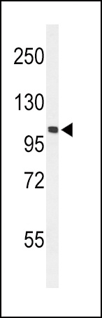
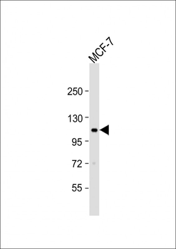
Western Blot at 1:1000 dilution + MCF-7 whole cell lysate Lysates/proteins at 20 ug per lane.
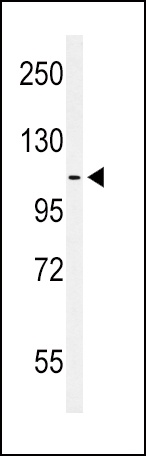
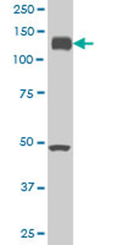
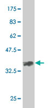
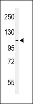
Western blot analysis of hEPHA2-T45 in MCF-7 cell line lysates (35 ug/lane)
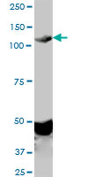
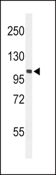
Western blot analysis of hEPHA2-T45 in mouse NIH-3T3 cell line lysates (35 ug/lane)
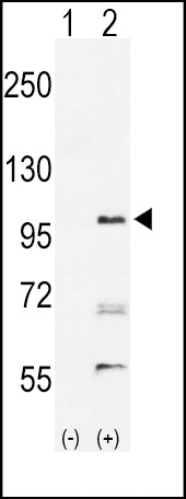
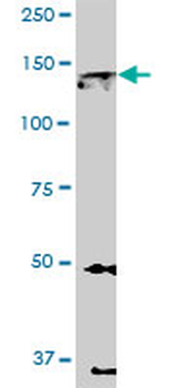
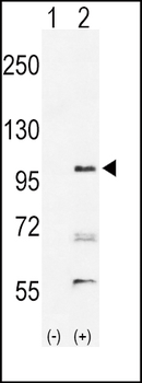
Western blot analysis of EPHA2 using rabbit polyclonal hEPHA2-T45 using 293 cell lysates (2 ug/lane) either nontransfected (Lane 1) or transiently transfected with the EPHA2 gene (Lane 2)
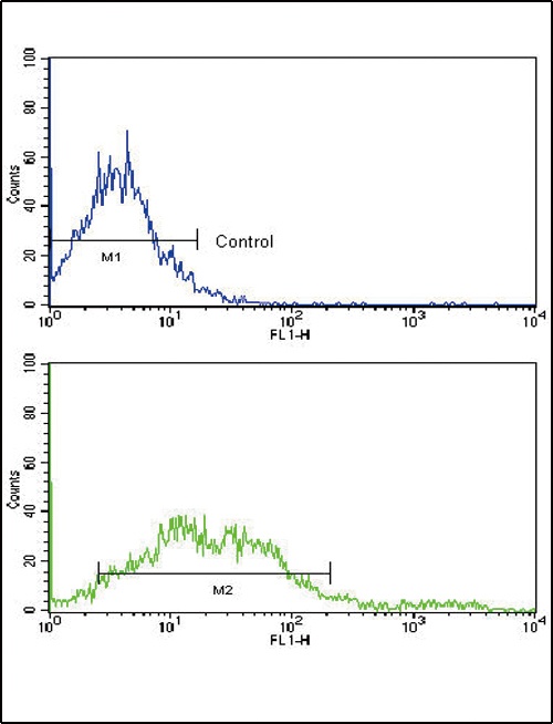
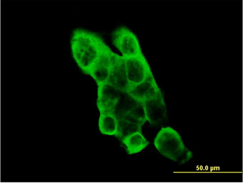
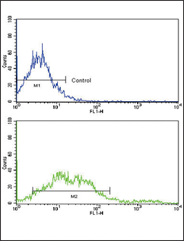
Flow cytometric analysis of NCI-H292 cells using EphA2 Antibody (N-term) (bottom histogram) compared to a negative control cell (top histogram). FITC-conjugated goat-anti-rabbit secondary antibodies were used for the analysis.
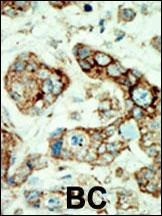
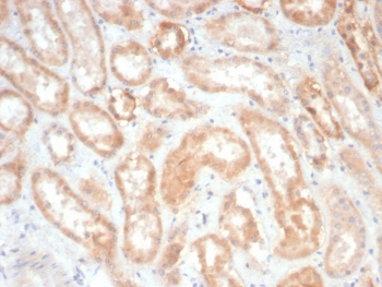
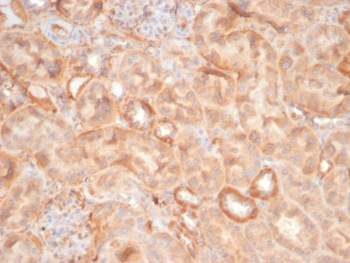
Formalin-fixed and paraffin-embedded human cancer tissue reacted with the primary antibody, which was peroxidase-conjugated to the secondary antibody, followed by AEC staining. BC = breast carcinoma; HC = hepatocarcinoma.
- Item 1 of 5
EPHA2 monoclonal antibody (M02), clone 1E3 [orb2293275]
ELISA, IF, WB
Human
Mouse
Monoclonal
Unconjugated
100 μg - Item 1 of 6
EphA2 Antibody (N-term) [orb1929104]
FC, IHC-P, WB
Human, Mouse
Rabbit
Polyclonal
Unconjugated
100 μl, 50 μl - Item 1 of 2
Anti-Human Ephrin Type A receptor 2 [1C1] [orb348860]
ELISA, FC, IF, WB
Human, Mouse
Human
Monoclonal
Unconjugated
0.2 mg - Item 1 of 2
Anti-Human Ephrin Type A receptor 2 [1C1] [orb348887]
ELISA, FC, IF, WB
Human, Mouse
Human
Monoclonal
Unconjugated
0.2 mg - Item 1 of 4

![Anti-Human Ephrin Type A receptor 2 [1C1]](/images//pub/media/catalog/product/NewWebsite/35/orb348860_1.png)
![Anti-Human Ephrin Type A receptor 2 [1C1]](/images/pub/media/catalog/product/NewWebsite/35/orb348860_2.png)
![Anti-Human Ephrin Type A receptor 2 [1C1]](/images//pub/media/catalog/product/NewWebsite/35/orb348887_1.png)
![Anti-Human Ephrin Type A receptor 2 [1C1]](/images/pub/media/catalog/product/NewWebsite/35/orb348887_2.png)