You have no items in your shopping cart.
Cart summary
Item 1 of 6
Item 1 of 6
ANXA2 Antibody
Catalog Number: orb1263234

| Catalog Number | orb1263234 |
|---|---|
| Category | Antibodies |
| Description | ANXA2 Antibody |
| Target | ANXA2 |
| Clonality | Polyclonal |
| Isotype | Rabbit Ig |
| Conjugation | Unconjugated |
| Reactivity | Human, Mouse |
| Predicted Reactivity | Bovine, Porcine, Rat |
| Form/Appearance | Liquid |
| Concentration | batch dependent |
| Buffer/Preservatives | Supplied in PBS with 0.09% (W/V) sodium azide. |
| Purification | This antibody is purified through a protein A column, followed by peptide affinity purification. |
| Immunogen | This ANXA2 antibody is generated from rabbits immunized with a KLH conjugated synthetic peptide between 19-49 amino acids from the N-terminal region of human ANXA2. |
| UniProt ID | P07355 |
| MW | 39 kDa |
| Tested applications | IHC-P, WB |
| Application notes | For IHC-P starting dilution is: 1:25For WB starting dilution is: 1:1000 |
| Antibody Type | Primary Antibody |
| Storage | Maintain refrigerated at 2-8°C for up to 2 weeks. For long term storage store at -20°C in small aliquots to prevent freeze-thaw cycles. |
| Alternative names | Annexin A2, Annexin II, Annexin-2, Calpactin I hea Read more... |
| Note | For research use only |
| NCBI | P07355 |
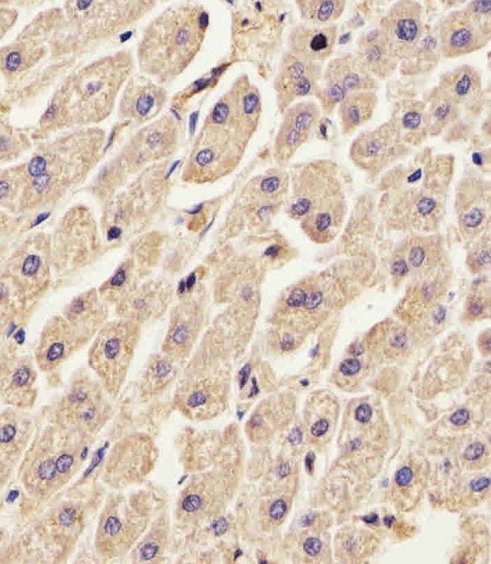
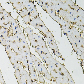
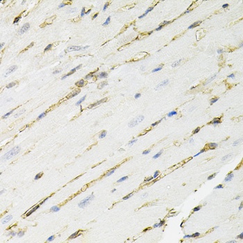
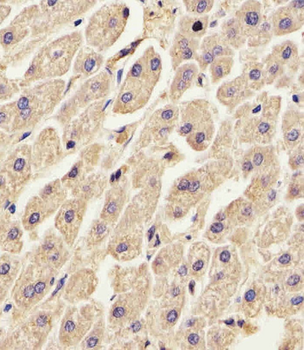
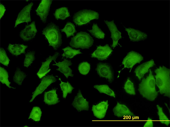
Antibody staining ANXA2 in Human liver tissue sections by Immunohistochemistry (IHC-P - paraformaldehyde-fixed, paraffin-embedded sections).
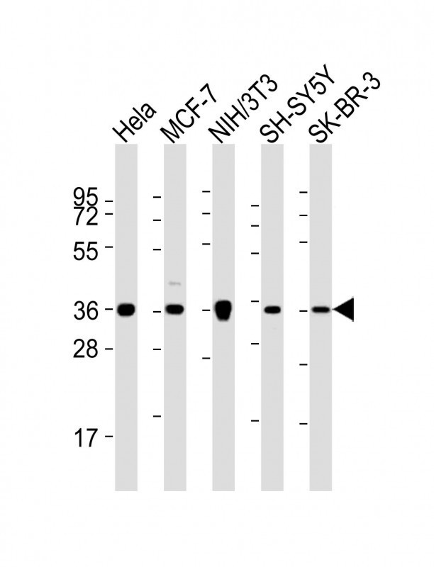
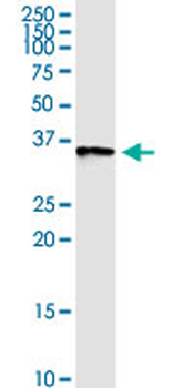
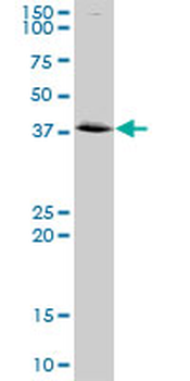
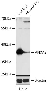
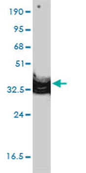
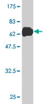
Western Blot at 1:2000 dilution Lane 1: Hela whole cell lysates Lane 2: MCF-7 whole cell lysates Lane 3: NIH/3T3 whole cell lysates Lane 4: SH-SY5Y whole cell lysates Lane 5: SK-BR-3 whole cell lysates Lysates/proteins at 20 ug per lane.

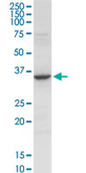
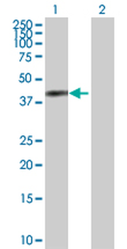
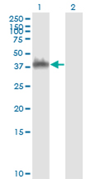
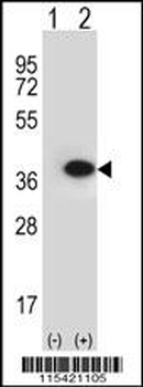
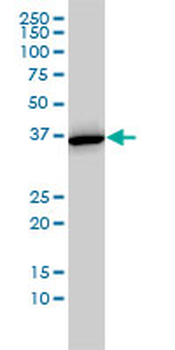
Western blot analysis of ANXA2 using rabbit polyclonal ANXA2 Antibody using 293 cell lysates (2 ug/lane) either nontransfected (Lane 1) or transiently transfected (Lane 2) with the ANXA2 gene.

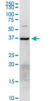
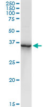
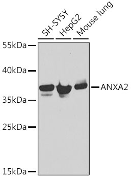
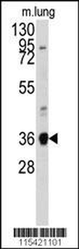
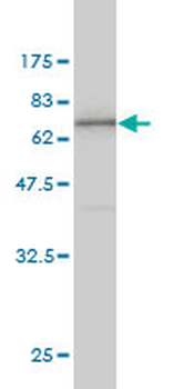
Western blot analysis of anti-ANXA2 Antibody in mouse lung tissue lysates (35 ug/lane).

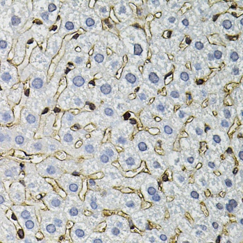
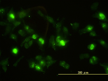
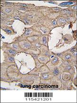
Formalin-fixed and paraffin-embedded human lung carcinoma tissue reacted with ANXA2 antibody, which was peroxidase-conjugated to the secondary antibody, followed by DAB staining.

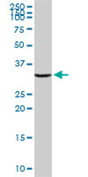
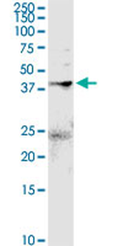
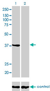
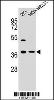
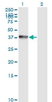
Western blot analysis in 293, MDA-MB231 cell line lysates (35 ug/lane).
- Item 1 of 8
ANXA2 purified MaxPab rabbit polyclonal antibody (D01P) [orb2296062]
WB
Human, Mouse
Rabbit
Polyclonal
Unconjugated
100 μg - Item 1 of 6
- Item 1 of 5
ANXA2 monoclonal antibody (M01), clone 3E8-B6 [orb2296061]
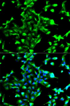
ELISA, IF, WB
Human
Mouse
Monoclonal
Unconjugated
100 μg - Item 1 of 6
ANXA2 Antibody (N-term) [orb1929277]
IHC-P, WB
Porcine, Rat, Sheep
Human, Mouse
Rabbit
Polyclonal
Unconjugated
100 μl, 50 μl - Item 1 of 4
ANXA2 monoclonal antibody (M02), clone 1G7 [orb2296060]
ELISA, IF, WB
Human
Mouse
Monoclonal
Unconjugated
100 μg