You have no items in your shopping cart.
Description
Research Area
Immunology & Inflammation
Images & Validation
−Item 1 of 2
| Tested Applications | FC, ICC |
|---|---|
| Reactivity | Human |
| Application Notes |
Key Properties
−| Clonality | Monoclonal |
|---|---|
| Isotype | Mouse IgG2b kappa |
| Clone No. | TB3 |
| Target | CD3 |
| Purification | Purified antibody is conjugated with biotin LC-NHS ester under optimum conditions and unconjugated antibody and free biotin are removed by size-exclusion chromatography. |
| Conjugation | Biotin |
Storage & Handling
−| Storage | Store at 2-8°C. Do not freeze. |
|---|---|
| Buffer/Preservatives | Phosphate buffered saline (PBS), pH 7.4, 15 mM sodium azide |
| Concentration | 1 mg/ml |
| Expiration Date | 12 months from date of receipt. |
| Disclaimer | For research use only |
Alternative Names
−CD3E, T3E, TCRE
Similar Products
−- Item 1 of 1
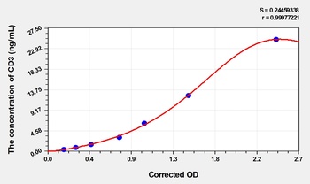
Mouse Cluster of Differentiation 3 (CD3) ELISA Kit [orb1817292]
Mouse
0.4-25 ng/mL
0.14 ng/mL
48 T, 96 T - Item 1 of 1
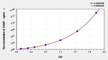
Human T-cell surface glycoprotein CD3 zeta Chain (CD247) ELISA Kit [orb1173602]
Human
0.16-10 ng/mL
0.059 ng/mL
48 T, 96 T - Item 1 of 1
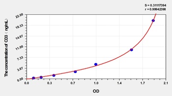
Human Cluster of Differentiation 3 (CD3) ELISA Kit [orb1173609]
Human
0.32-20 ng/mL
0.128 ng/mL
96 T, 48 T - Item 1 of 2
- Item 1 of 2
Quality Guarantee
Explore bioreagents carefree to elevate your research. All our products are rigorously tested for performance. If a product does not perform as described on its datasheet, our scientific support team will provide expert troubleshooting, a prompt replacement, or a refund. For full details, please see our Terms & Conditions and Buying Guide. Contact us at [email protected].
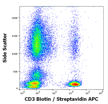
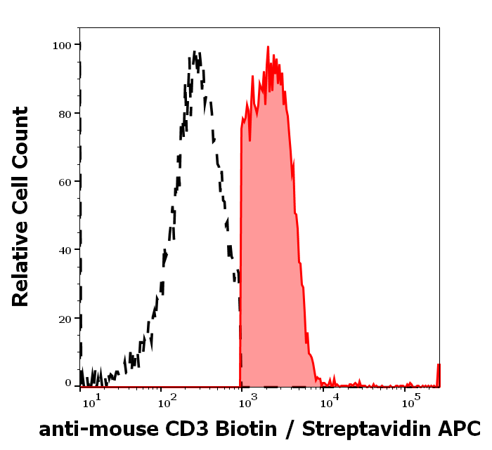
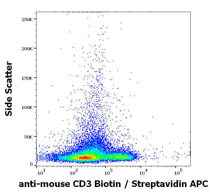
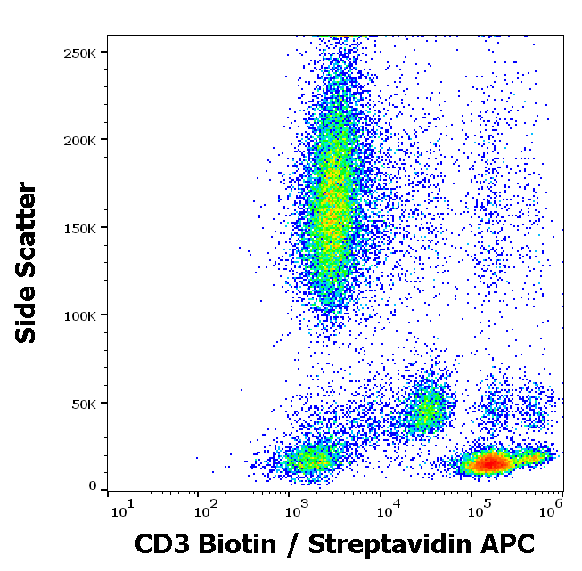
Flow cytometry surface staining pattern of human peripheral whole blood stained using anti-human CD3 (TB3) biotin antibody (concentration in sample 0.56 µg/ml, Streptavidin APC).
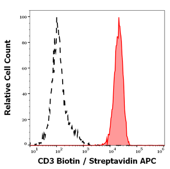
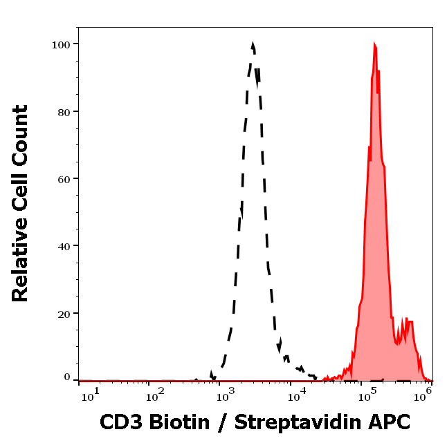
Separation of human CD3 positive lymphocytes (red-filled) from neutrophil granulocytes (black-dashed) in flow cytometry analysis (surface staining) of human peripheral whole blood stained using anti-human CD3 (TB3) biotin antibody (concentration in sample 0.56 µg/ml, Streptavidin APC).
Documents Download
Datasheet
Product Information
Request a Document
Protocol Information
FC
Flow Cytometry
ICC
Immunocytochemistry
CD3 Antibody (Biotin) (orb2704159)

- 0.0
Based on 0 reviews
Participating in our Biorbyt product reviews program enables you to support fellow scientists by sharing your firsthand experience with our products.
Login to Submit a ReviewAvailable Sizes
Select a size below
Free Secondary Antibody (20 ul)0/0
Please add an antibody product to your cart first.