You have no items in your shopping cart.
Cart summary
Item 1 of 9
Item 1 of 9
Alpha Synuclein Pre-formed Fibrils (Type 1)
Catalog Number: orb1822365

| Catalog Number | orb1822365 |
|---|---|
| Category | Proteins |
| Description | Human Recombinant Alpha Synuclein Pre-formed Fibrils (Type 1) |
| Target | Alpha Synuclein |
| Tag | No tag |
| Reactivity | Human |
| Concentration | 2 mg/ml or 5 mg/ml |
| Buffer/Preservatives | PBS |
| Purity | > 95% |
| Protein Sequence | MDVFMKGLSK AKEGVVAAAE KTKQGVAEAA GKTKEGVLYV GSKTKEGVVH GVATVAEKTK EQVTNVGGAV VTGVTAVAQK TVEGAGSIAA ATGFVKKDQL GKNEEGAPQE GILEDMPVDP DNEAYEMPSE EGYQDYEPEA |
| Protein Length | Full Length |
| UniProt ID | P37840 |
| MW | ~14.46 kDa |
| Tested applications | In vitro, In vivo, SDS-PAGE, WB |
| Application notes | Certified > 95% pure using SDS-PAGE analysis. Low endotoxin < 5 EU/mL @ 2mg/mL. |
| Expression System | E. coli |
| Source | Recombinant |
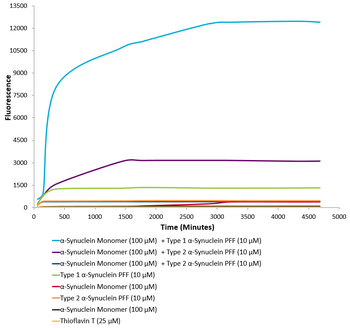
| Biological Activity | Endogenous alpha-synuclein phosphorylation. 100 µM alpha synuclein protein monomer seeded with 10 uM alpha synuclein protein PFF in 25 µM Thioflavin T (PBS pH 7.4, 100 µl reaction volume) generated a fluorescence intensity of 13,000 Relative Fluorescence Units after incubation at 37°C with shaking at 600 rpm. Fluorescence was measured by excitation at 450 nm and emission at 485 nm on a Molecular Devices Gemini XPS microplate reader. |
| Storage | -80°C |
| Alternative names | Alpha synuclein PFFs, Alpha synuclein aggregates, Read more... |
| Note | For research use only |

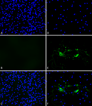
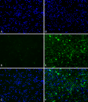
Primary rat hippocampal neurons show lewy body inclusion formation when treated with Type 1 Alpha Synuclein Protein Pre-formed Fibrils at 4 μg/ml (D-F), but not when treated with Type 2 Alpha Synuclein Protein Pre-formed Fibrils at 4 μg/ml (A-C). Tissue: Primary hippocampal neurons. Species: Sprague-Dawley rat. Fixation: 4% formaldehyde from PFA. Primary Antibody: Mouse anti-pSer129 Antibody at 1:1000 24 hours at 4°C. Secondary Antibody: FITC Goat Anti-Mouse (green) at 1:700 for 1 hours at RT. Counterstain: Hoechst (blue) nuclear stain at 1:4000 for 1 hour at RT. Localization: Lewy body inclusions. Magnification: 20x.
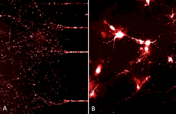
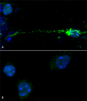
ATTO633 fluorescently-labelled alpha synuclein PFFs were taken up, transported into the soma, and induced alpha synuclein aggregation in mouse neurocortical primary cells. (A) Neurites filled with fluorescently-labelled alpha synuclein seeds in a microfluidic co-culture system after 24 hours. (B) Alpha synuclein seeds within the soma and neurites of mouse neurocortical primary cells after 24 hours.
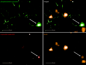
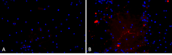
Confocal imaging shows NeuN+ (mature) primary cortical neurons filled with ATTO633 fluorescently-labelled alpha synuclein PFFs. ATTO-633 alpha synuclein PFFs seed endogenous alpha synculein phosphorylation after 7 days.
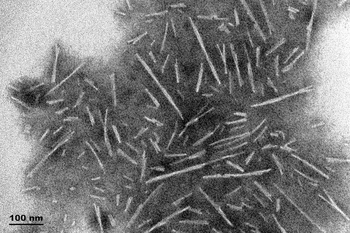
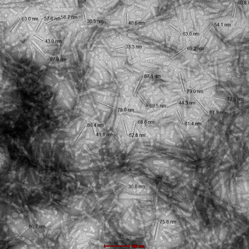
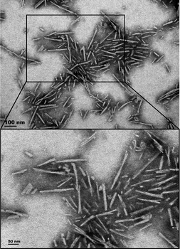
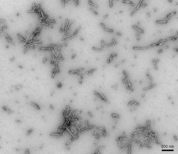
TEM of Type 1 Alpha Synuclein Pre-formed Fibrils (PFFs)
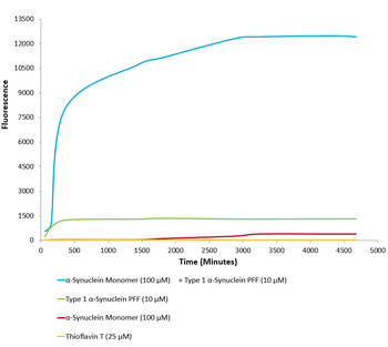
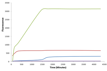
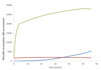
Type 1 alpha synuclein Pre-formed Fibrils seed the formation of new alpha synuclein fibrils from the pool of alpha synuclein monomers. Thioflavin T is a fluorescent dye that binds to beta sheet-rich structures, such as those in alpha synuclein fibrils. Upon binding, the emission spectrum of the dye experiences a red-shift, and increased fluorescence intensity. Thioflavin T emission curves show increased fluorescence (correlated to alpha synuclein protein aggregation) over time when 10 μM of Type 1 alpha synuclein Pre-formed Fibrils is combined with 100 μM of alpha synuclein monomer, as compared to Type 1 alpha synuclein Pre-formed Fibrils alone and alpha synuclein monomer alone. Thioflavin T ex = 450 nm, em = 485 nm.
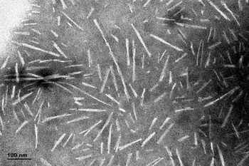
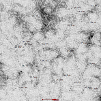
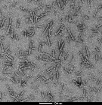
TEM of Type 1 Alpha Synuclein Pre-formed Fibrils (PFFs)
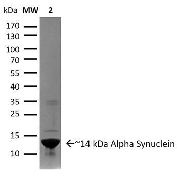
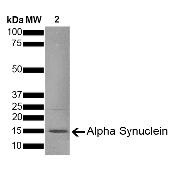
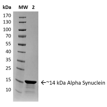
SDS-PAGE of ~14 kDa Type 1 Human Recombinant Alpha Synuclein Protein Pre-formed Fibrils. Lane 1: Molecular Weight Ladder (MW). Lane 2: Type 1 Alpha Synuclein Protein Pre-formed Fibrils.

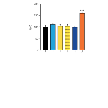
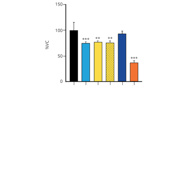
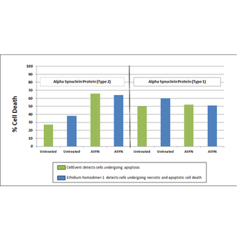
Toxicity results comparing Active Human Recombinant Alpha Synuclein Pre-formed Fibrils (Type 2) and Active Human Recombinant Alpha Synuclein Pre-formed Fibrils (Type 1). Data was graphed after live cell imaging results were obtained using the following procedure: After 8 days in vitro, primary rat mixed cortical neuron cells were washed with 1X PBS and treated with 500 μg/ml of Type 1 and Type 2 Alpha Synuclein Proteins for 20 hours at 37?C. Following treatements, cells were washed with 2X PBS and incubated with a staining solution (2.0 μM Cell Event + 2.5 μM Ethidium homodimer + 2.5 μg/ml Hoechst 33342 in sterile HBSS) for 30 minutes at 37?C. The addition of the Type 2 Alpha Synuclein Proteins resulted in a significant increase in cell death.
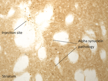
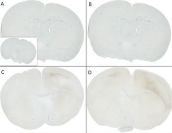
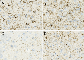
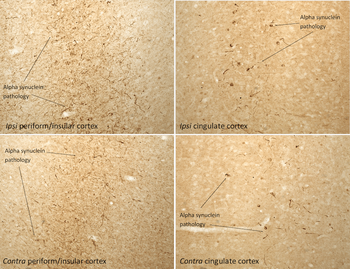
Immunohistochemistry analysis of rat brain injected with Type 1 human alpha synuclein PFFs. Species: Female Sprague-Dawley Rat. Rat was injected with 16μg Type 1 human alpha synuclein PFFs in each of 2 injection sites: AP+1.6, ML+2.4, DV-4.2 from skull; and AP-1.4, ML+0.2, DV-2.8 from skull. 30-days post-injection. Fixation: Saline perfusion followed by 4% PFA fixation for 48 hrs. Primary antibody: rabbit monoclonal anti-pSer129 alpha synuclein. Secondary Antibody: Biotin-SP Donkey Anti-Rabbit IgG (H+L) at 1:500 for 2 hours in cold room with shaking. ABC signal amplification, DAB staining. Magnification: 20X. Alpha synuclein pathology is seen in the striatum close to an injection site.
- Item 1 of 9
Alpha Synuclein Pre-formed Fibrils (Type 1) [orb1822362]
In vitro, In vivo, SDS-PAGE, WB
> 95%
~14.46 kDa
Recombinant
100 μg - Item 1 of 8
Alpha Synuclein A53T Mutant Pre-formed Fibrils (Type 1) [orb1822360]
In vitro, In vivo, SDS-PAGE, WB
> 95%
~14.46 kDa
Recombinant
100 μg - Item 1 of 7
Alpha Synuclein Pre-formed Fibrils (Type 2) [orb1822369]
In vitro, In vivo, SDS-PAGE, WB
> 95%
~14.46 kDa
Recombinant
100 μg