You have no items in your shopping cart.
Cart summary
Item 1 of 5
Item 1 of 5
AKR1B1 Antibody
Catalog Number: orb1264639

| Catalog Number | orb1264639 |
|---|---|
| Category | Antibodies |
| Description | AKR1B1 Antibody |
| Target | AKR1B1 |
| Clonality | Polyclonal |
| Isotype | Rabbit Ig |
| Conjugation | Unconjugated |
| Reactivity | Human |
| Form/Appearance | Liquid |
| Concentration | batch dependent |
| Buffer/Preservatives | Supplied in PBS with 0.09% (W/V) sodium azide. |
| Immunogen | This AKR1B1 antibody is generated from rabbits immunized with a KLH conjugated synthetic peptide between 102-135 amino acids from the Central region of human AKR1B1. |
| UniProt ID | P15121 |
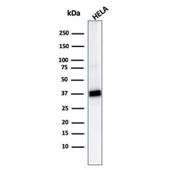
| MW | 36 kDa |
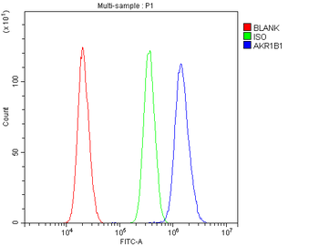
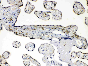
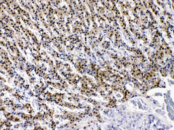
| Tested applications | IF, IHC-P, WB |
| Application notes | For WB starting dilution is: 1:1000For IF starting dilution is: 1:10~50For IHC-P starting dilution is: 1:10~50 |
| Antibody Type | Primary Antibody |
| Storage | Maintain refrigerated at 2-8°C for up to 2 weeks. For long term storage store at -20°C in small aliquots to prevent freeze-thaw cycles. |
| Alternative names | Aldose reductase, AR, Aldehyde reductase, Aldo-ket Read more... |
| Note | For research use only |
| NCBI | P15121 |
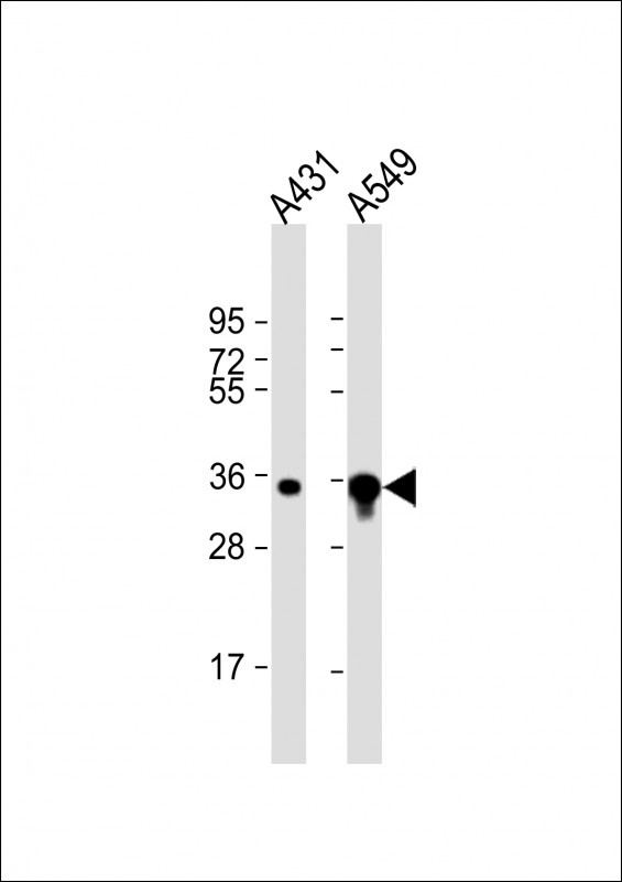
Western Blot at 1:8000 dilution Lane 1: A431 whole cell lysate Lane 2: A549 whole cell lysate Lysates/proteins at 20 ug per lane.
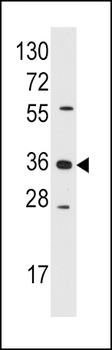
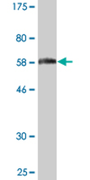
Western blot analysis of anti-AKR1B1 Pab in Jurkat cell line lysates (35 ug/lane)

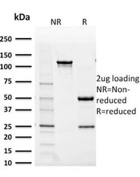
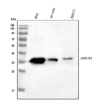
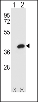
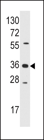
Western blot analysis of AKR1B1 using rabbit polyclonal AKR1B1 Antibody using 293 cell lysates (2 ug/lane) either nontransfected (Lane 1) or transiently transfected (Lane 2) with the AKR1B1 gene.

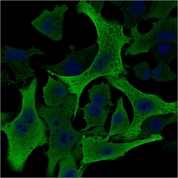
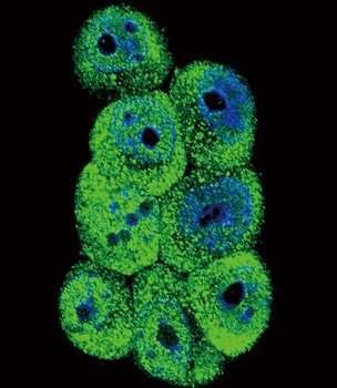
Confocal immunofluorescent analysis of AKR1B1 Antibody with 293 cell followed by Alexa Fluor 488-conjugated goat anti-rabbit lgG (green). DAPI was used to stain the cell nuclear (blue).
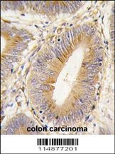
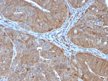
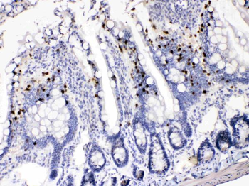
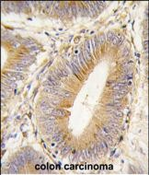
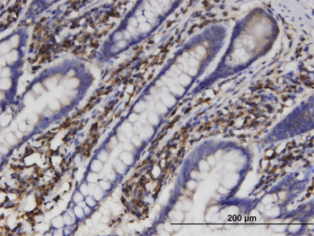
Formalin-fixed and paraffin-embedded human colon carcinoma tissue reacted with AKR1B1 antibody, which was peroxidase-conjugated to the secondary antibody, followed by DAB staining.
- Item 1 of 5
- Item 1 of 5
AKR1B1 Antibody / Aldose reductase [orb2641482]
IF, IHC-P, WB
Human
Mouse
Monoclonal
Unconjugated
100 μg - Item 1 of 5
Anti-AKR1B1 Antibody [orb381037]
FC, IHC, WB
Human, Mouse, Rat
Rabbit
Polyclonal
Unconjugated
10 μg, 100 μg - Item 1 of 5
- Item 1 of 3
AKR1B1 monoclonal antibody (M03), clone 2D12 [orb2296167]
ELISA, IHC-P, WB
Human
Mouse
Monoclonal
Unconjugated
100 μg