You have no items in your shopping cart.
Cart summary
Item 1 of 7
Item 1 of 7
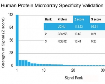
UCHL1 Antibody
Catalog Number: orb1264982

| Catalog Number | orb1264982 |
|---|---|
| Category | Antibodies |
| Description | UCHL1 Antibody |
| Target | UCHL1 |
| Clonality | Polyclonal |
| Isotype | Rabbit Ig |
| Conjugation | Unconjugated |
| Reactivity | Human, Mouse, Rat |
| Predicted Reactivity | Bovine, Monkey, Porcine |
| Form/Appearance | Liquid |
| Concentration | batch dependent |
| Buffer/Preservatives | Supplied in PBS with 0.09% (W/V) sodium azide. |
| Immunogen | This UCHL1 antibody is generated from rabbits immunized with a KLH conjugated synthetic peptide between 187-216 amino acids from the C-terminal region of human UCHL1. |
| UniProt ID | P09936 |
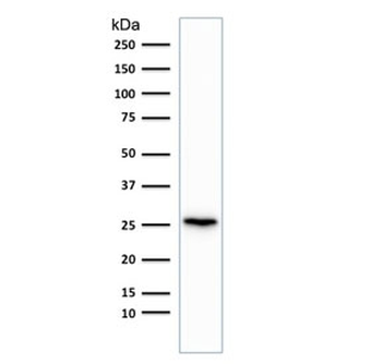
| MW | 25 kDa |
| Tested applications | FC, IF, IHC-P, WB |
| Application notes | For IHC-P starting dilution is: 1:25For WB starting dilution is: 1:1000For FACS starting dilution is: 1:10~50For IF starting dilution is: 1:10~50 |
| Antibody Type | Primary Antibody |
| Storage | Maintain refrigerated at 2-8°C for up to 2 weeks. For long term storage store at -20°C in small aliquots to prevent freeze-thaw cycles. |
| Alternative names | Ubiquitin carboxyl-terminal hydrolase isozyme L1, Read more... |
| Note | For research use only |
| NCBI | P09936 |
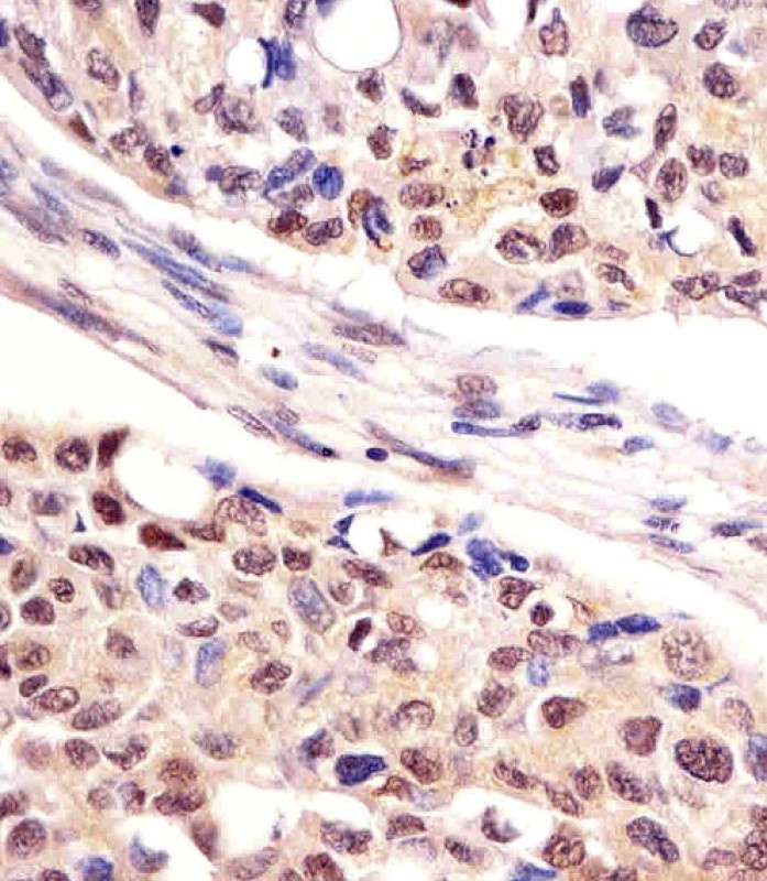
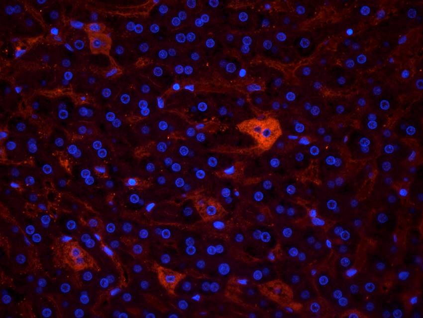
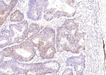
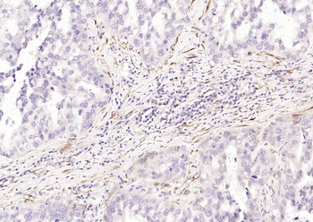
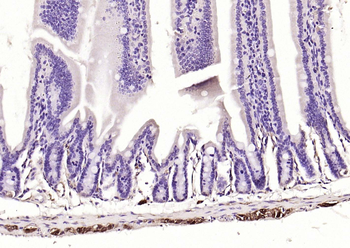
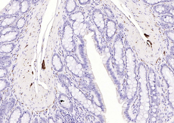
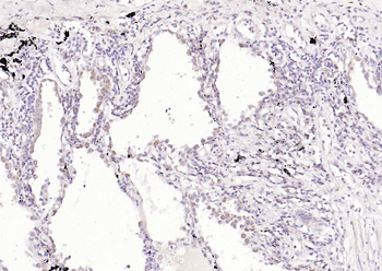
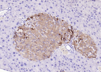
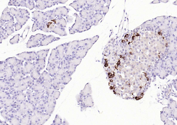
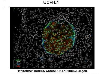
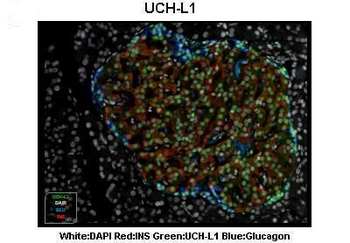
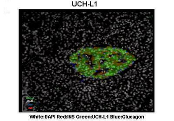
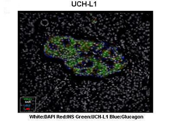
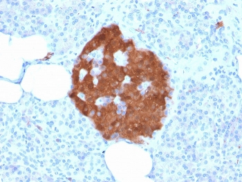
Antibody staining UCHL1 in human lung adenocarcinoma tissue sections by Immunohistochemistry (IHC-P - paraformaldehyde-fixed, paraffin-embedded sections).
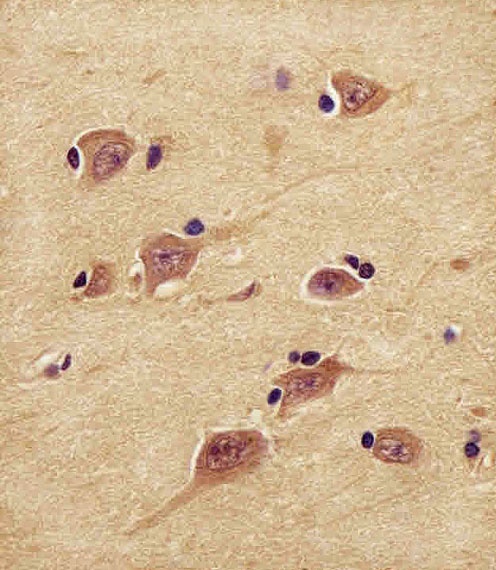
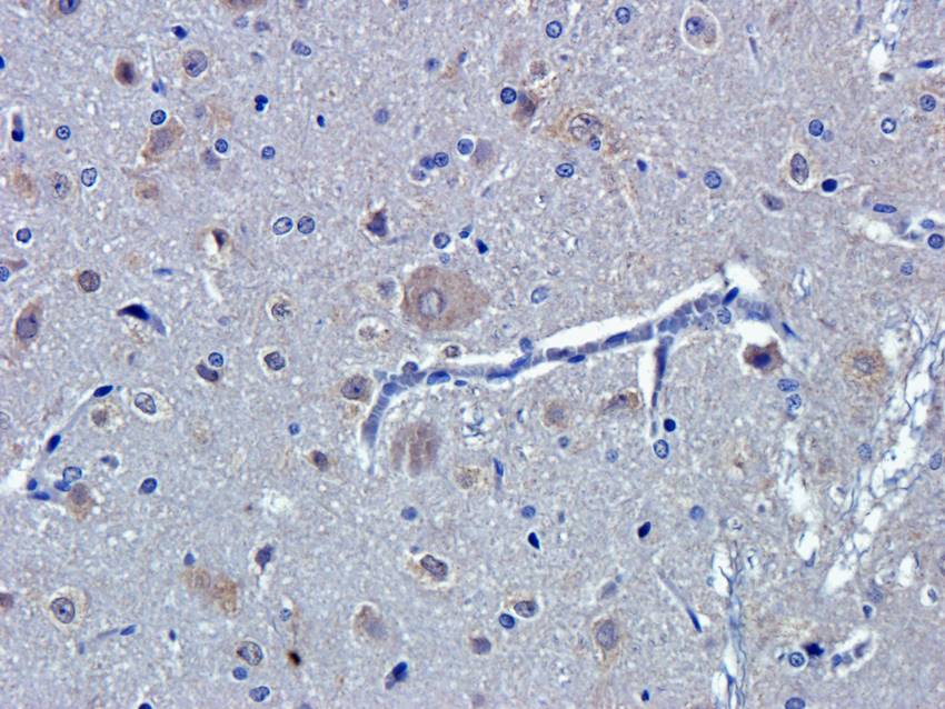
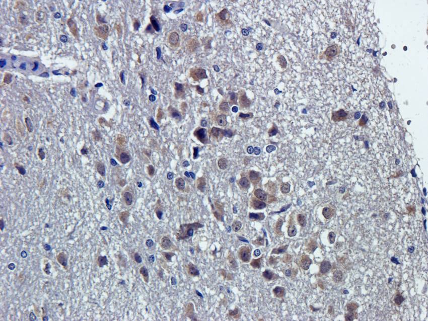
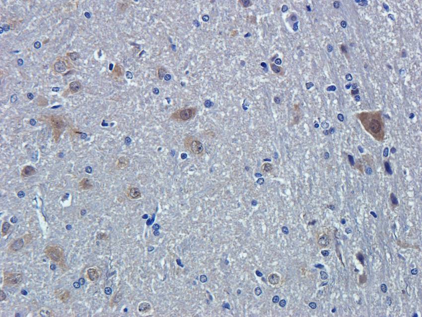
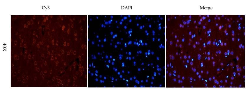
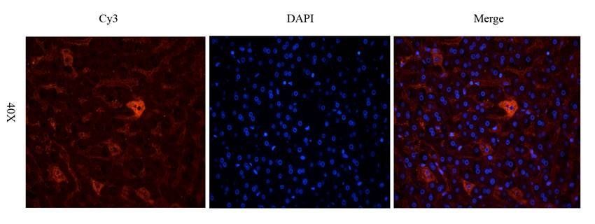
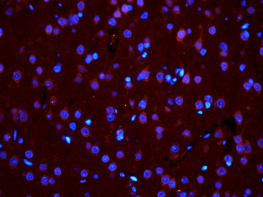
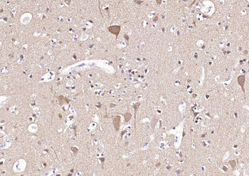
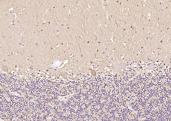
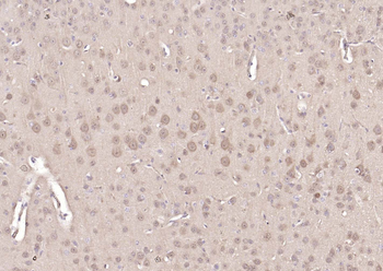
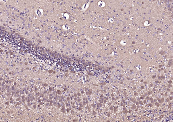
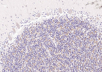
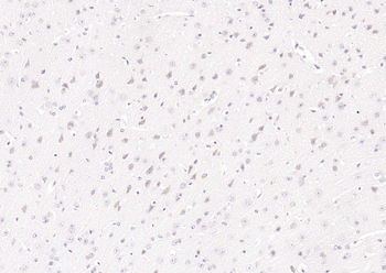
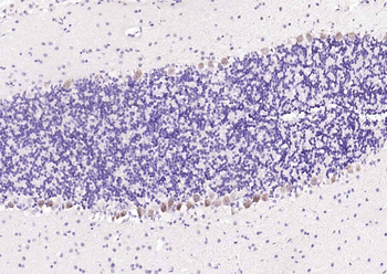
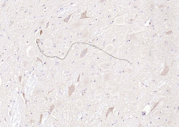
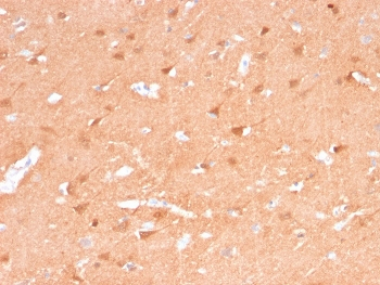
Antibody staining UCHL1 in human brain tissue sections by Immunohistochemistry (IHC-P - paraformaldehyde-fixed, paraffin-embedded sections).
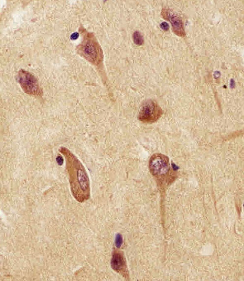
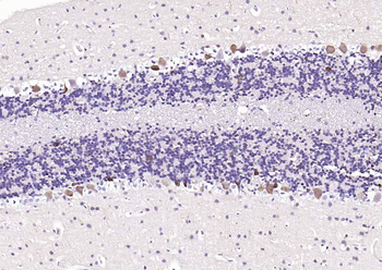
Antibody staining UCHL1 in human brain tissue sections by Immunohistochemistry (IHC-P - paraformaldehyde-fixed, paraffin-embedded sections).
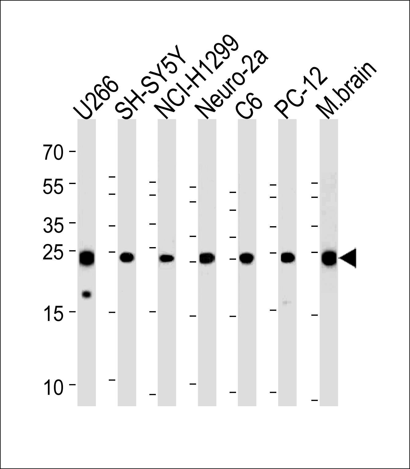
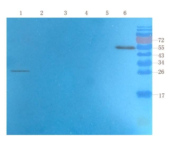
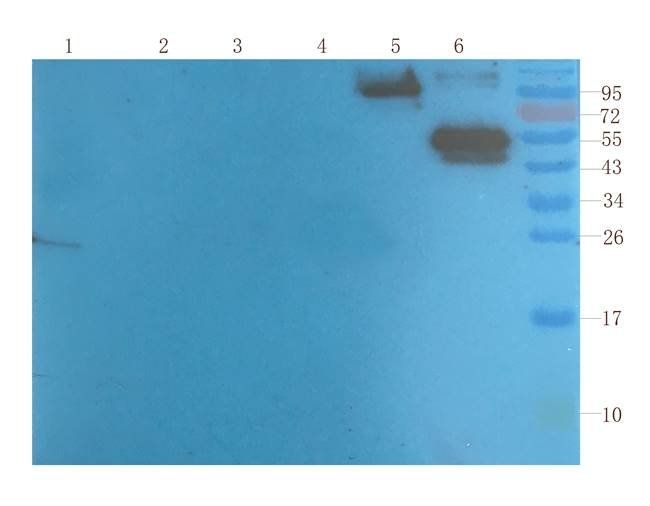
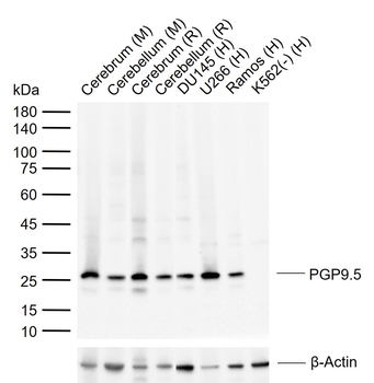
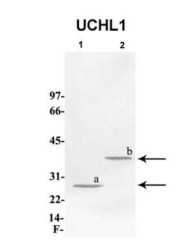
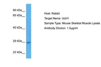
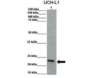
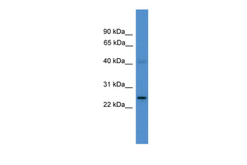
Western blot analysis in U266, SH-SY5Y, NCI-H1299, mouse Neuro-2a, rat C6, PC-12 cell line and mouse brain tissue lysates (35 ug/lane).
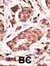
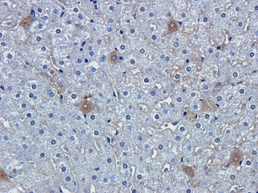
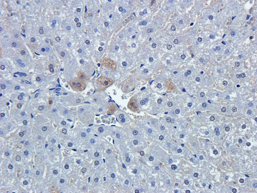
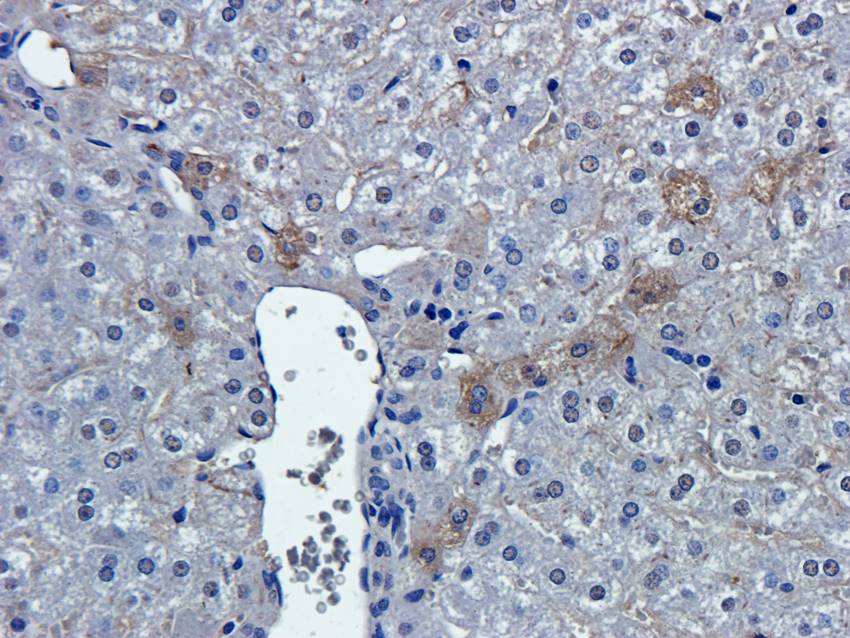
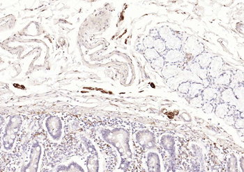
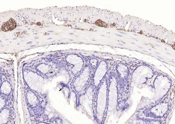
Formalin-fixed and paraffin-embedded human cancer tissue reacted with the primary antibody, which was peroxidase-conjugated to the secondary antibody, followed by DAB staining. BC = breast carcinoma; HC = hepatocarcinoma.
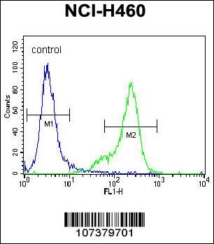
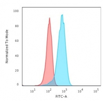
Flow cytometric analysis of NCI-H460 cells (right histogram) compared to a negative control cell (left histogram). FITC-conjugated goat-anti-rabbit secondary antibodies were used for the analysis.
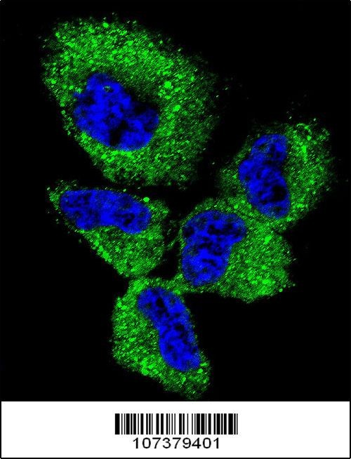
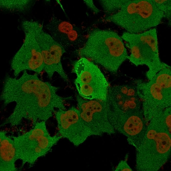
Confocal immunofluorescent analysis of UCHL1 Antibody with NCI-H460 cell followed by Alexa Fluor 488-conjugated goat anti-rabbit lgG (green).DAPI was used to stain the cell nuclear (blue).
- Item 1 of 12
PGP9.5 antibody [orb6713]
ELISA, ICC, IF, IHC-P, WB
Human, Mouse, Rat
Rabbit
Polyclonal
Unconjugated
100 μg - Item 1 of 14
PGP9.5 Mouse Monoclonal Antibody [orb2563495]
IF, IHC-Fr, IHC-P, WB
Human, Mouse, Rat
Human, Mouse, Rat
Mouse
Monoclonal
Unconjugated
200 μg, 100 μl, 200 μl, 50 μl - Item 1 of 7
PGP9.5 Rabbit Polyclonal Antibody [orb500977]
IF, IHC-Fr, IHC-P
Equine, Guinea pig, Porcine
Bovine, Human, Mouse, Rat
Rabbit
Polyclonal
Unconjugated
100 μl, 200 μl, 50 μl - Item 1 of 8
UCHL1 Rabbit Polyclonal Antibody [orb331031]
IHC, WB
Bovine, Canine, Equine, Guinea pig, Rabbit, Zebrafish
Human, Mouse, Rat
Rabbit
Polyclonal
Unconjugated
100 μl - Item 1 of 7