You have no items in your shopping cart.
Cart summary
Item 1 of 4
Item 1 of 4
TUBB3 Antibody
Catalog Number: orb1672015
| Catalog Number | orb1672015 |
|---|---|
| Category | Antibodies |
| Description | TUBB3 Antibody |
| Clonality | Recombinant |
| Clone Number | S11B |
| Tested applications | ELISA, WB |
| Reactivity | Human |
| Isotype | IgG lambda |
| Immunogen | Human MBP (microtubule-binding protein). |
| Concentration | batch dependent |
| Conjugation | Unconjugated |
| Target | TUBB3 |
| UniProt ID | Q13509 |
| Storage | Store at 4°C for up to 3 months. For longer storage, aliquot and store at -20°C. |
| Buffer/Preservatives | PBS with 0.02% Proclin 300. |
| Alternative names | TUBB Read more... |
| Note | For research use only |
| Expiration Date | 12 months from date of receipt. |
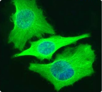
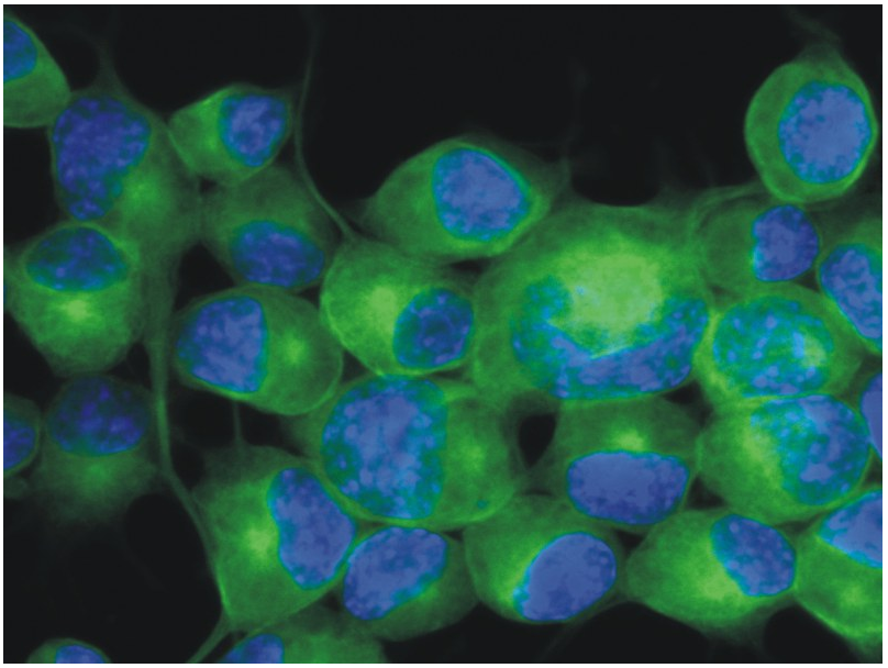
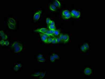
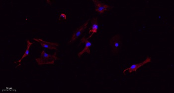
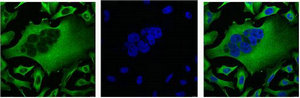
Immunofluoresence staining of fixed HeLa cells with anti-Beta-tubulin antibody S11B. Immunofluorescence analysis of paraformaldehyde fixed HeLa cells- permewith 0.15% Triton stained with the chimeric r version of S11B (orb1672015) at 10 ug/ml for 1h followed by Alexa Fluor® 488 secondary antibody (1 ug/ml)- showing cytoplasmic staining. The nuclear stain is DAPI (blue). Panels show from left-right- top-bottom orb1672015- DAPI- merged channels and a negative control. The negative control was stained with unimmunized r followed by Alexa Fluor® 488 secondary antibody.
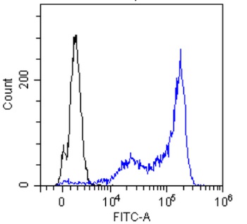
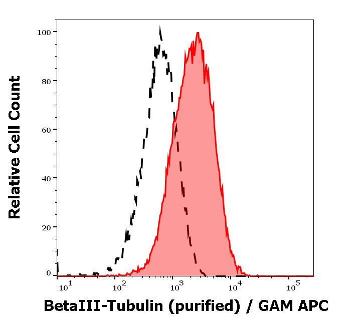
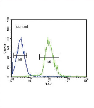
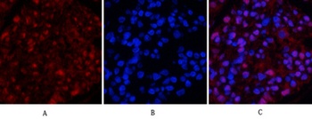
Flow-cytometry using the anti-Beta Tubulin S11B. MCF-7 (A) and HeLa (B) cells were stained with unimmunized r antibody (black line) or the rmeric version of S11B (orb1672015 - blue line) at a concentration of 10 ug/ml for 30 mins at RT. After washing- bound antibody was detected using anti-r JK (FITC-conjugate) antibody (129936) at 2 ug/ml and cells analyzed on a FACSCanto flow-cytometer.
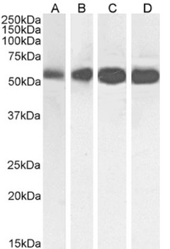
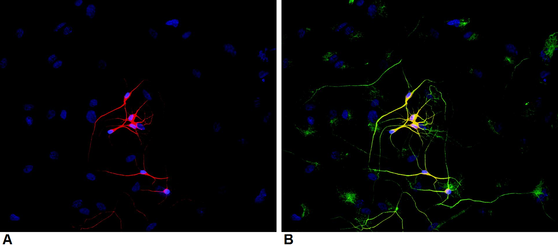
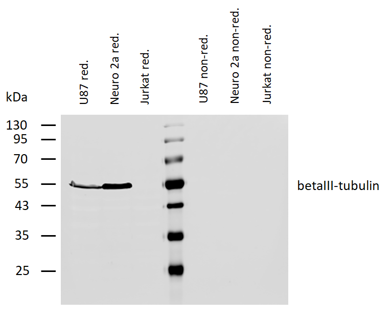
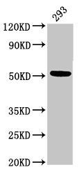
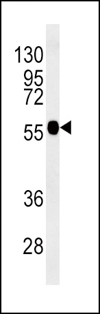
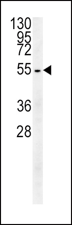
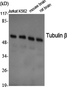
Western Blot using anti-Beta-Tubulin antibody S11B. HeLa (A)- A431 (B)- HEK293 (C) and MCF-7 (D) cell lysate samples (35 ug protein in RIPA buffer) were resolved on a 10% SDS PAGE gel and blots probed with the chimeric rsion of S11B (orb1672015) at 0.01 ug/ml before detection using an anti-rondary antibody. A primary incubation of 1h was used and protein was detected by chemiluminescence. The expected band size for Beta-Tubulin is ~54kDa. orb1672015 successfully detected human Beta-Tubulin in HeLa- A431- HEK293 and MCF-7 cell lysate samples.
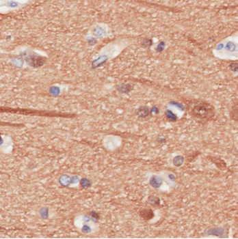
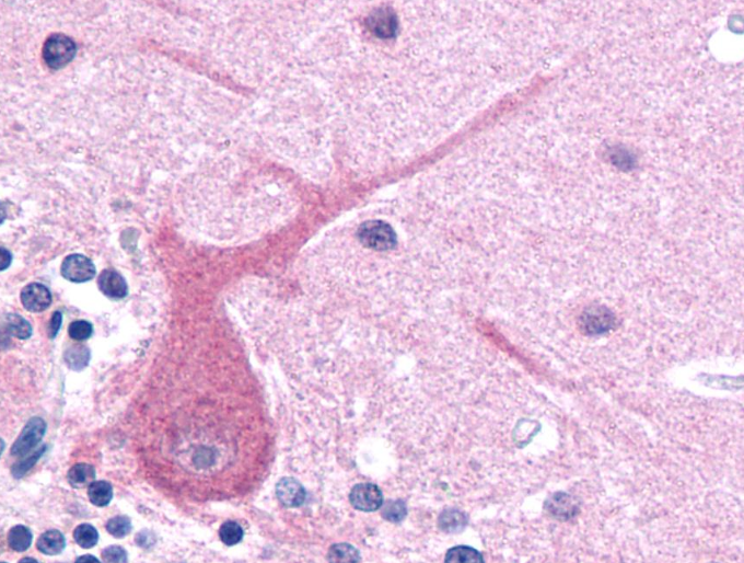
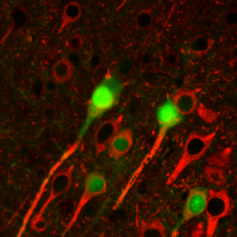
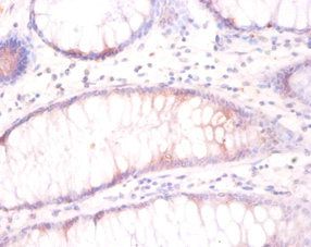
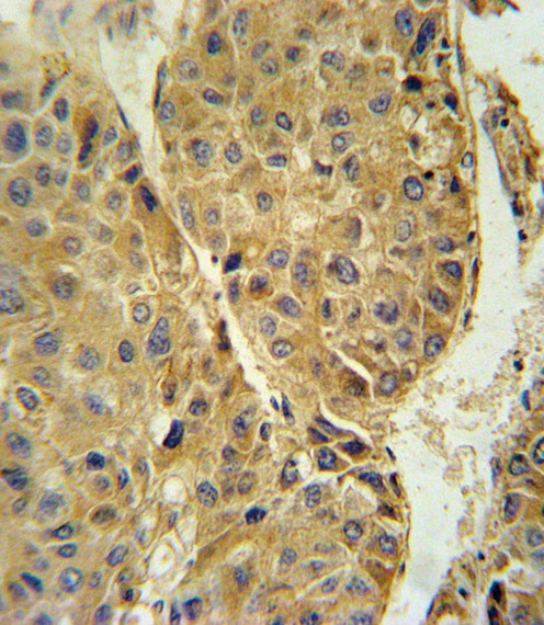
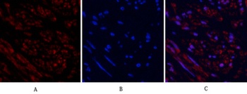
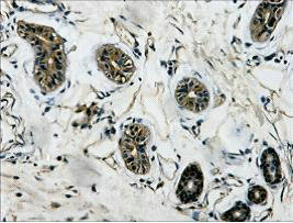
Immunohistochemical staining of human cerebral cortex tissue using anti-Beta Tubulin antibody. S11B Anti-Beta Tubulin staining of paraffin embedded human cerebral cortex tissue using the rmeric version of S11B (orb1672015). Antigen retreival was acheived by microwaving in citrate buffer (pH6)- followed by blocking with protein block serum-free buffer. Primary antibody incubation with orb1672015 was carried out at 4 ug/ml for 30 minutes. Samples were then incubated with an anti-r HRP secondary antibody for 20 mins followed by Ddiaminobenzidine- and counter-staining with haemotoxylin. Staining of neuronal cell bodies and their processes may be observed. Recommended concentration- 2-4 ug/ml.
- Item 1 of 6
Tubulin beta 3 antibody [orb44544]
FC, ICC, IHC-P, WB
Canine, Human, Mouse, Porcine, Rat
Monoclonal
Unconjugated
0.1 mg - Item 1 of 5
Tubulin beta 3 antibody [orb241857]
ELISA, IF, IHC, WB
Human
Rabbit
Polyclonal
Unconjugated
50 μg, 100 μg - Item 1 of 5
TUBB3 Antibody [orb1264082]
FC, IHC-P, WB
Bovine, Gallus, Monkey, Rat
Human, Mouse
Rabbit
Polyclonal
Unconjugated
400 μl - Item 1 of 4
Tubulin beta antibody [orb766531]
ELISA, IF, IHC-P, WB
Bovine, Human, Mouse, Porcine, Rat
Rabbit
Polyclonal
Unconjugated
50ul, 100ul - Item 1 of 3
Tubulin beta 3 antibody [orb688348]
ELISA, IHC, WB
Canine, Gallus, Hamster, Human, Insect, Monkey, Mouse, Rabbit, Rat, Sheep, Yeast
Mouse
Monoclonal
Unconjugated
100 μg, 50 μg

Submit a review
Filter by Rating

- 5 stars

- 4 stars
- 3 stars
- 2 stars
- 1 stars