You have no items in your shopping cart.
Cart summary
Item 1 of 6
Item 1 of 6
TrkA-pY791 Antibody
Catalog Number: orb1263001

| Catalog Number | orb1263001 |
|---|---|
| Category | Antibodies |
| Description | TrkA-pY791 Antibody |
| Target | NTRK1 |
| Clonality | Polyclonal |
| Isotype | Rabbit Ig |
| Conjugation | Unconjugated |
| Reactivity | Human, Mouse |
| Form/Appearance | Liquid |
| Concentration | batch dependent |
| Buffer/Preservatives | Supplied in PBS with 0.09% (W/V) sodium azide. |
| Purification | This antibody is purified through a protein A column, followed by peptide affinity purification. |
| Immunogen | This TrkA antibody is generated from rabbits immunized with a KLH conjugated synthetic peptide between 769-796 amino acids from human TrkA. |
| UniProt ID | P04629 |
| MW | 87 kDa |
| Tested applications | FC, IHC-P, WB |
| Application notes | For FACS starting dilution is: 1:25For IHC-P starting dilution is: 1:25For WB starting dilution is: 1:1000 |
| Antibody Type | Primary Antibody |
| Storage | Maintain refrigerated at 2-8°C for up to 2 weeks. For long term storage store at -20°C in small aliquots to prevent freeze-thaw cycles. |
| Alternative names | High affinity nerve growth factor receptor, Neurot Read more... |
| Note | For research use only |
| NCBI | P04629 |
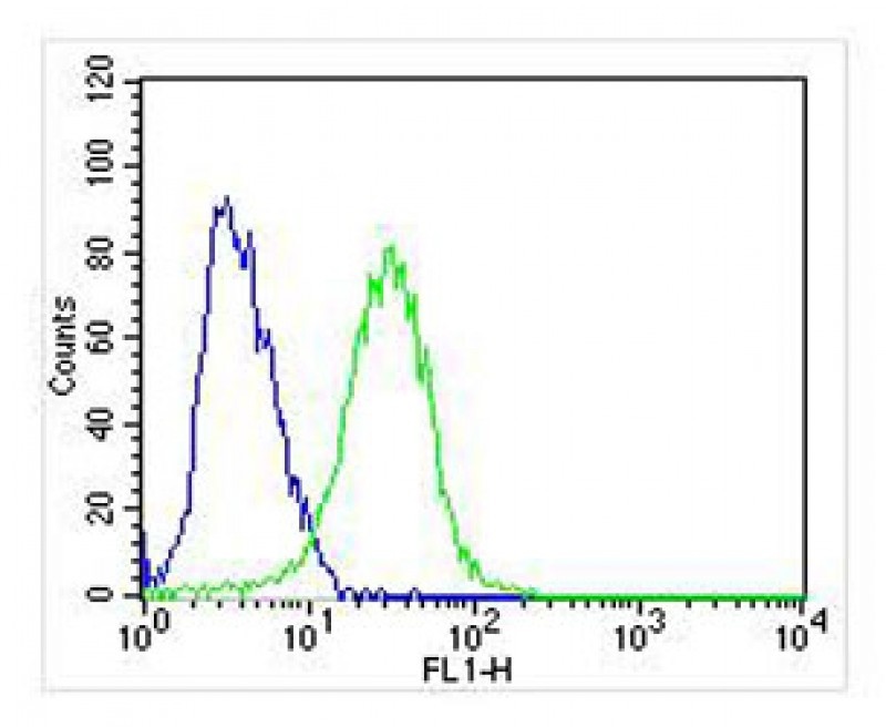
Overlay histogram showing SH-SY5Y cells stained with Antibody (green line). The cells were fixed with 2% paraformaldehyde (10 min). The cells were then icubated in 2% bovine serum albumin to block non-specific protein-protein interactions followed by the antibody (1:25 dilution) for 60 min at 37°C. The secondary antibody used was Goat-Anti-Rabbit IgG, Conjugated Highly Cross-Adsorbed at 1/400 dilution for 40 min at 37°C. Isotype control antibody (blue line) was rabbit IgG (1ug/1x10^6 cells) used under the same conditions. Acquisition of > 10000 events was performed.

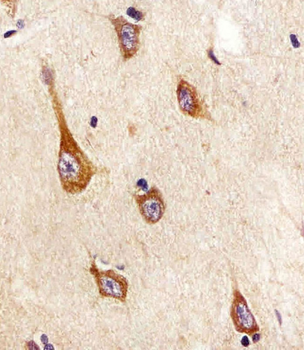
Antibody staining TrkA in human brain tissue sections by Immunohistochemistry (IHC-P - paraformaldehyde-fixed, paraffin-embedded sections).
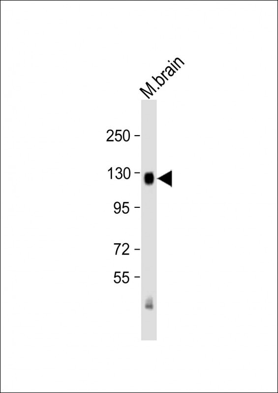
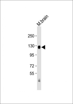
Western Blot at 1:2000 dilution + mouse brain lysate Lysates/proteins at 20 ug per lane.
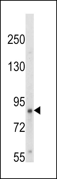
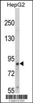
Western blot analysis of hTrkA-pY791 in HepG2 cell line lysates (35 ug/lane)
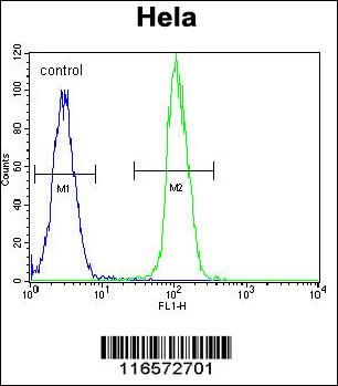
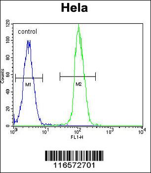
Flow cytometric analysis of Hela cells (right histogram) compared to a negative control cell (left histogram). FITC-conjugated goat-anti-rabbit secondary antibodies were used for the analysis.
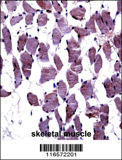
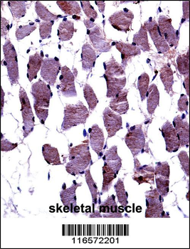
TrkA-pY791 Antibody immunohistochemistry analysis in formalin fixed and paraffin embedded human skeletal muscle followed by peroxidase conjugation of the secondary antibody and DAB staining.
- Item 1 of 6
TrkA-pY791 Antibody [orb1928982]
FC, IHC-P, WB
Human, Mouse
Rabbit
Polyclonal
Unconjugated
100 μl, 50 μl