You have no items in your shopping cart.
Cart summary
Item 1 of 4
Item 1 of 4
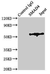
SMAD4 Antibody
Catalog Number: orb1262961

| Catalog Number | orb1262961 |
|---|---|
| Category | Antibodies |
| Description | SMAD4 Antibody |
| Species/Host | Rabbit |
| Clonality | Polyclonal |
| Tested applications | FC, IF, IHC-P, WB |
| Predicted Reactivity | Bovine, Mouse, Porcine, Rat |
| Reactivity | Human |
| Isotype | Rabbit Ig |
| Immunogen | This SMAD4 antibody is generated from rabbits immunized with a KLH conjugated synthetic peptide between 400-428 amino acids from the C-terminal region of human SMAD4. |
| Antibody Type | Primary Antibody |
| Concentration | batch dependent |
| Form/Appearance | Liquid |
| Conjugation | Unconjugated |
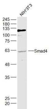
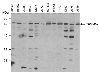
| MW | 60 kDa |
| Target | SMAD4 |
| UniProt ID | Q13485 |
| NCBI | Q13485 |
| Storage | Maintain refrigerated at 2-8°C for up to 2 weeks. For long term storage store at -20°C in small aliquots to prevent freeze-thaw cycles. |
| Buffer/Preservatives | Supplied in PBS with 0.09% (W/V) sodium azide. |
| Alternative names | Mothers against decapentaplegic homolog 4, MAD hom Read more... |
| Note | For research use only |
| Application notes | For WB starting dilution is: 1:1000For IHC-P starting dilution is: 1:10~50For IF starting dilution is: 1:10~50For FACS starting dilution is: 1:10~50 |
| Expiration Date | 12 months from date of receipt. |
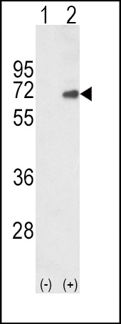
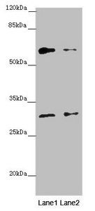
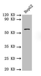
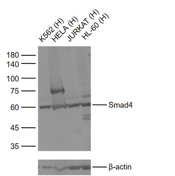
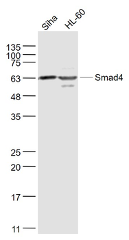
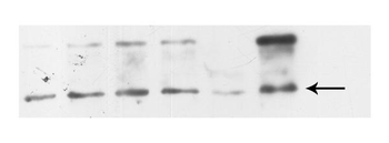
Western blot analysis of SMAD4 using rabbit polyclonal SMAD4 Antibody using 293 cell lysates (2 ug/lane) either nontransfected (Lane 1) or transiently transfected with the SMAD4 gene (Lane 2).
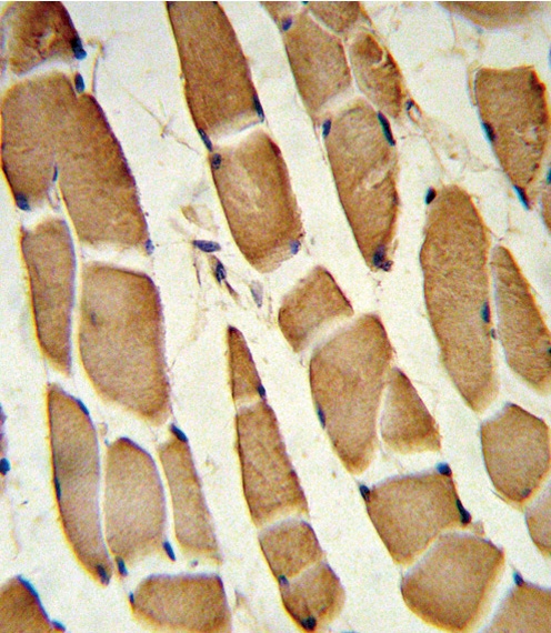
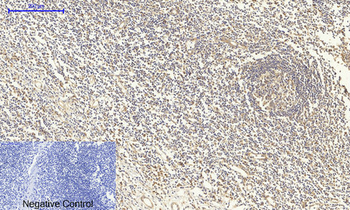
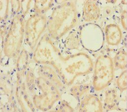
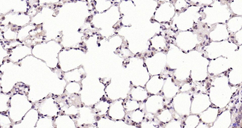
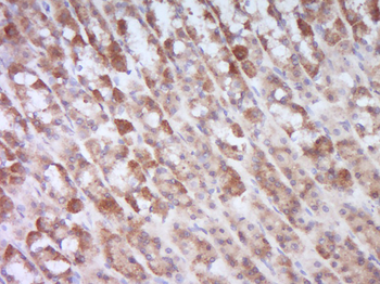
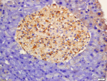
Formalin-fixed and paraffin-embedded human skeletal muscle reacted with SMAD4 Antibody, which was peroxidase-conjugated to the secondary antibody, followed by DAB staining.
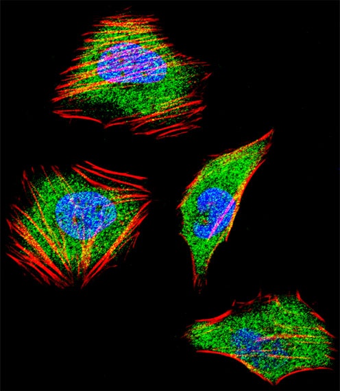
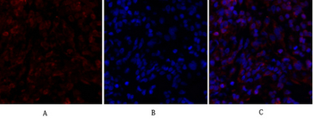
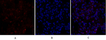
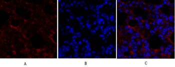
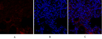
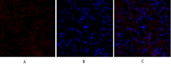
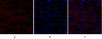
Fluorescent confocal image of Hela cell stained with SMAD4 Antibody.Hela cells were fixed with 4% PFA (20 min), permeabilized with Triton X-100 (0.1%, 10 min), then incubated with SMAD4 primary antibody (1:25). For secondary antibody, Alexa Fluor 488 conjugated donkey anti-rabbit antibody (green) was used (1:400).Cytoplasmic actin was counterstained with Alexa Fluor 555 (red) conjugated Phalloidin (7 units/ml). Nuclei were counterstained with DAPI (blue) (10 ug/ml, 10 min). SMAD4 immunoreactivity is localized to Cytoplasm and Nucleus significantly.
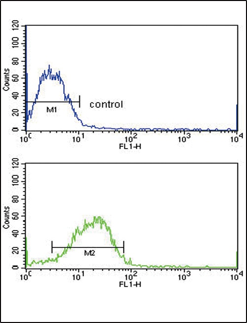
Flow cytometric analysis of MCF-7 cells (bottom histogram) compared to a negative control cell (top histogram). FITC-conjugated goat-anti-rabbit secondary antibodies were used for the analysis.
- Item 1 of 7
Smad4 Polyclonal Antibody [orb1412146]
IF, IHC-P, WB
Human, Mouse, Rat
Rabbit
Polyclonal
Unconjugated
100 μl - Item 1 of 5
- Item 1 of 3
Smad4 Rabbit Polyclonal Antibody [orb526558]
WB
Bovine, Canine, Equine, Gallus, Mouse, Porcine, Rabbit, Rat, Sheep, Zebrafish
Human
Rabbit
Polyclonal
Unconjugated
50 μl, 100 μl, 200 μl - Item 1 of 3
Smad4 Rabbit Polyclonal Antibody [orb11385]
WB
Bovine, Canine, Equine, Gallus, Mouse, Porcine, Rabbit, Rat, Sheep, Zebrafish
Human
Rabbit
Polyclonal
Unconjugated
100 μl, 200 μl, 50 μl - Item 1 of 4
SMAD4 Rabbit Polyclonal Antibody [orb574011]
WB
Bovine, Canine, Equine, Goat, Guinea pig, Mouse, Rabbit, Rat, Sheep
Human
Rabbit
Polyclonal
Unconjugated
100 μl