You have no items in your shopping cart.
Cart summary
Item 1 of 6
Item 1 of 6
SEPT9 Antibody
Catalog Number: orb1263937

| Catalog Number | orb1263937 |
|---|---|
| Category | Antibodies |
| Description | SEPT9 Antibody |
| Target | SEPT9 |
| Clonality | Polyclonal |
| Isotype | Rabbit Ig |
| Conjugation | Unconjugated |
| Reactivity | Human |
| Form/Appearance | Liquid |
| Concentration | batch dependent |
| Buffer/Preservatives | Supplied in PBS with 0.09% (W/V) sodium azide. |
| Purification | This antibody is purified through a protein A column, followed by peptide affinity purification. |
| Immunogen | This SEPT9 antibody is generated from rabbits immunized with a KLH conjugated synthetic peptide between 57-85 amino acids from the C-terminal region of human SEPT9. |
| UniProt ID | Q9UHD8 |
| MW | 65 kDa |
| Tested applications | FC, IHC-P, WB |
| Application notes | For WB starting dilution is: 1:1000For IHC-P starting dilution is: 1:50~100For FACS starting dilution is: 1:10~50 |
| Antibody Type | Primary Antibody |
| Storage | Maintain refrigerated at 2-8°C for up to 2 weeks. For long term storage store at -20°C in small aliquots to prevent freeze-thaw cycles. |
| Alternative names | Septin-9, MLL septin-like fusion protein MSF-A, ML Read more... |
| Note | For research use only |
| NCBI | Q9UHD8 |

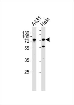
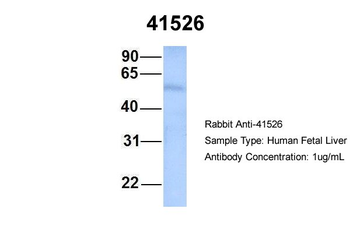
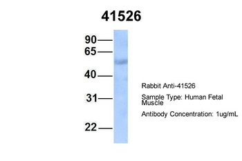
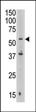
Western blot analysis of lysates from A431, Hela cell line (from left to right), using SEPT9 Antibody at 1:1000 at each lane.

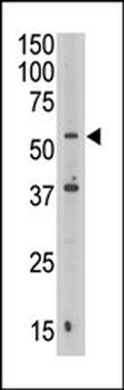
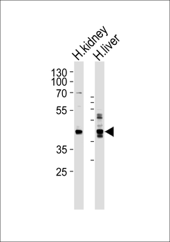
Western blot analysis of lysates from human kidney and liver tissue lysate (from left to right), using SEPT9 Antibody at 1:1000 at each lane.

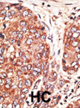
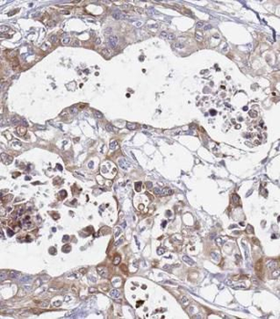
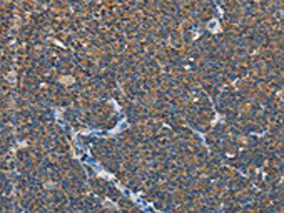
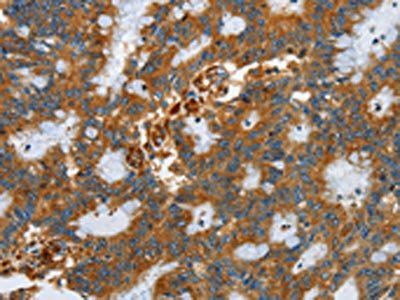
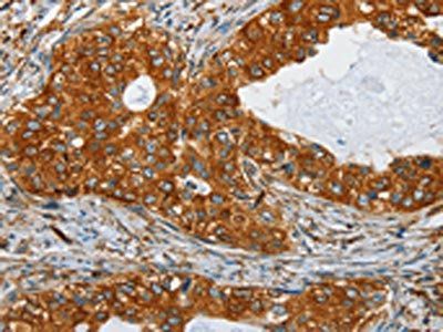
Formalin-fixed and paraffin-embedded human cancer tissue reacted with the primary antibody, which was peroxidase-conjugated to the secondary antibody, followed by AEC staining. BC = breast carcinoma; HC = hepatocarcinoma.
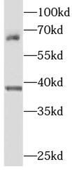
Antibody is used in Western blot to detect SEPT9 in Jurkat cell lysate.
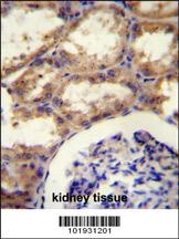
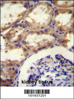
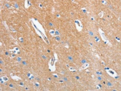
SEPT9 Antibody (A555) immunohistochemistry analysis in formalin fixed and paraffin embedded kidney tissue followed by peroxidase conjugation of the secondary antibody and DAB staining.This data demonstrates the use of SEPT9 Antibody (A555) for immunohistochemistry.
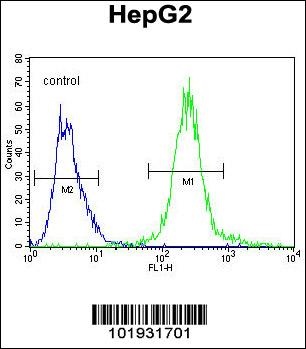
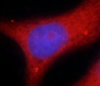
Flow cytometric analysis of HepG2 cells (right histogram) compared to a negative control cell (left histogram). FITC-conjugated goat-anti-rabbit secondary antibodies were used for the analysis.
- Item 1 of 6
- Item 1 of 4
SEPTIN9 Antibody [orb395566]
ELISA, FC, IF, IHC, IP, WB
Human, Mouse, Rat
Rabbit
Polyclonal
Unconjugated
50 μg, 100 μg - Item 1 of 3
SEPT9 Rabbit Polyclonal Antibody [orb325685]
WB
Bovine, Canine, Equine, Guinea pig, Mouse, Rabbit, Rat, Zebrafish
Human
Rabbit
Polyclonal
Unconjugated
100 μl - Item 1 of 2
- Item 1 of 2