You have no items in your shopping cart.
Cart summary
Item 1 of 5
Item 1 of 5
RBX1 Antibody
Catalog Number: orb1265034

| Catalog Number | orb1265034 |
|---|---|
| Category | Antibodies |
| Description | RBX1 Antibody |
| Target | RBX1 |
| Clonality | Polyclonal |
| Isotype | Rabbit Ig |
| Conjugation | Unconjugated |
| Reactivity | Human |
| Predicted Reactivity | C. elegans, Mouse |
| Form/Appearance | Liquid |
| Concentration | batch dependent |
| Buffer/Preservatives | Supplied in PBS with 0.09% (W/V) sodium azide. |
| Purification | This antibody is purified through a protein A column, followed by peptide affinity purification. |
| Immunogen | This RBX1 antibody is generated from a rabbit immunized with a KLH conjugated synthetic peptide between 74-108 amino acids from the C-terminal region of human RBX1. |
| UniProt ID | P62877 |
| MW | 12 kDa |
| Tested applications | IF, IHC-P, WB |
| Application notes | For WB starting dilution is: 1:1000For IHC-P starting dilution is: 1:25For IF starting dilution is: 1:25 |
| Antibody Type | Primary Antibody |
| Storage | Maintain refrigerated at 2-8°C for up to 2 weeks. For long term storage store at -20°C in small aliquots to prevent freeze-thaw cycles. |
| Alternative names | E3 ubiquitin-protein ligase RBX1, 632-, Protein ZY Read more... |
| Note | For research use only |
| NCBI | P62877 |
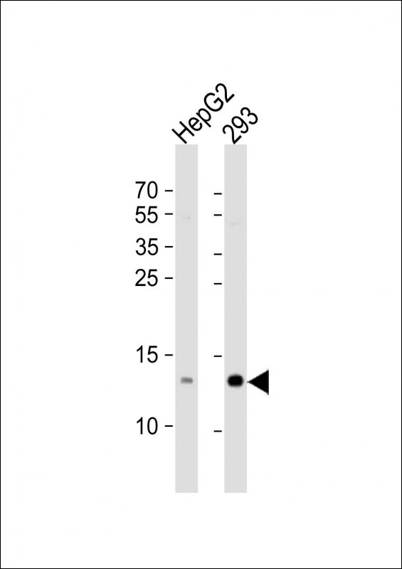
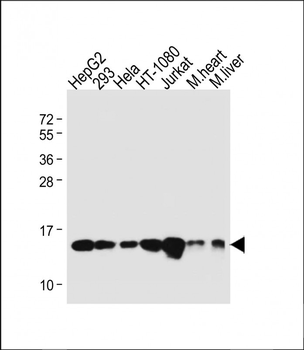
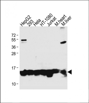
Western Blot at 1:1000 dilution Lane 1: HepG2 whole cell lysate Lane 2: 293 whole cell lysate Lysates/proteins at 20 ug per lane.

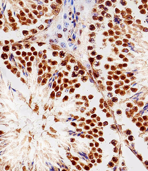
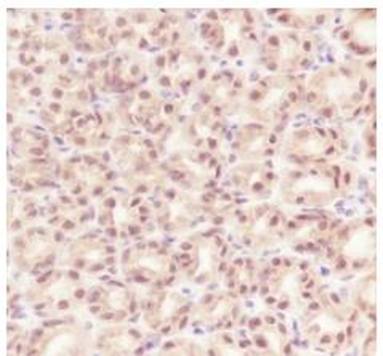
Immunohistochemical analysis of paraffin-embedded M. testis section using RBX1 Antibody. Antibody was diluted at 1:100 dilution. A peroxidase-conjugated goat anti-rabbit IgG at 1:400 dilution was used as the secondary antibody, followed by DAB staining.
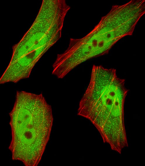
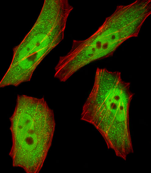
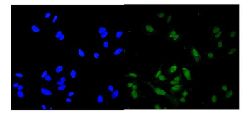
Fluorescent image of HeLa cells stained with RBX1 Antibody. Antibody was diluted at 1:25 dilution. An Alexa Fluor 488-conjugated goat anti-rabbit lgG at 1:400 dilution was used as the secondary antibody (green). Cytoplasmic actin was counterstained with Alexa Fluor 555 conjugated with Phalloidin (red).
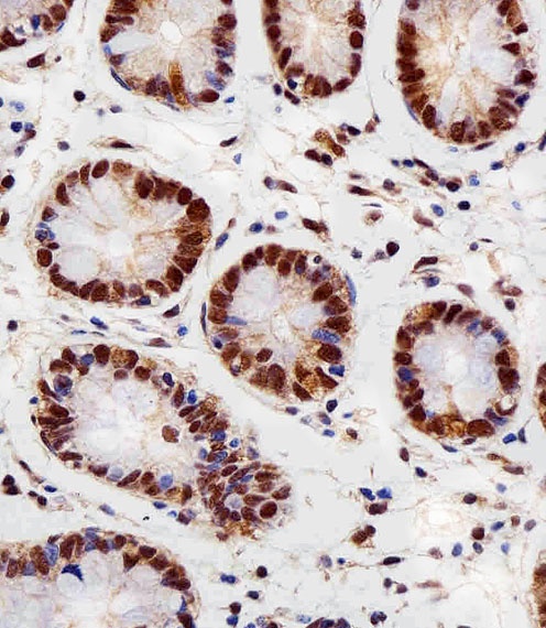
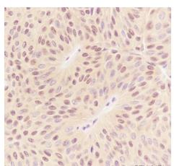
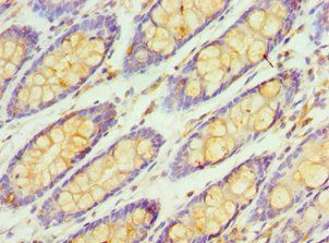
Immunohistochemical analysis of paraffin-embedded H. colon section using RBX1 Antibody. Antibody was diluted at 1:100 dilution. A peroxidase-conjugated goat anti-rabbit IgG at 1:400 dilution was used as the secondary antibody, followed by DAB staining.
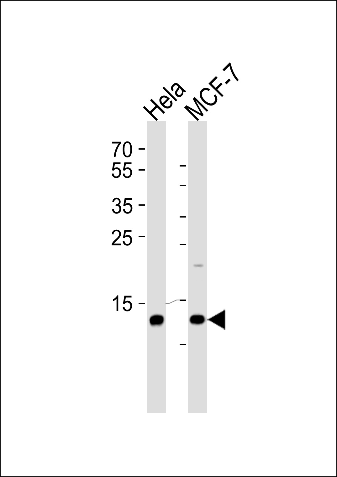
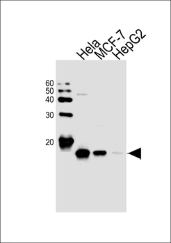
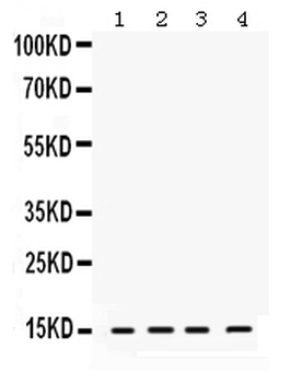
Western blot analysis of lysates from Hela, MCF-7 cell line (from left to right), using RBX1 Antibody at 1:1000 at each lane.
- Item 1 of 7
RBX1 Antibody (C-term) [orb1927189]
IF, IHC-P, WB
C. elegans
Human, Mouse
Rabbit
Polyclonal
Unconjugated
100 μl, 50 μl - Item 1 of 5
RBX1 Antibody (C-term) [orb1788447]
IF, IHC-P, WB
C. elegans, Mouse
Human
Rabbit
Polyclonal
Unconjugated
100 μl - Item 1 of 3
RBX1 Recombinant Rabbit Monoclonal Antibody [orb1499278]
ICC, IF, IHC-Fr, IHC-P, WB
Mouse
Human, Rat
Rabbit
Recombinant
Unconjugated
25 μl, 100 μl, 50 μl - Item 1 of 2
- Item 1 of 3
Anti-ROC1/RBX1 Antibody [orb334530]
FC, ICC, IF, WB
Human, Mouse, Rat
Rabbit
Polyclonal
Unconjugated
10 μg, 100 μg