You have no items in your shopping cart.
Cart summary
Item 1 of 7
Item 1 of 7
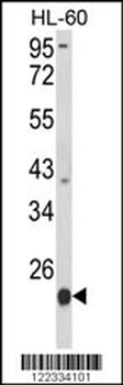
PSMB9 Antibody (C-term)
Catalog Number: orb1788016

| Catalog Number | orb1788016 |
|---|---|
| Category | Antibodies |
| Description | Purified Rabbit Polyclonal Antibody (Pab) |
| Target | This PSMB9 antibody is generated from a rabbit immunized with a KLH conjugated synthetic peptide between 205-239 amino acids from the C-terminal region of human PSMB9. |
| Clonality | Polyclonal |
| Species/Host | Rabbit |
| Isotype | Rabbit IgG |
| Conjugation | Unconjugated |
| Reactivity | Human |
| Form/Appearance | Purified polyclonal antibody supplied in PBS with 0.09% (W/V) sodium azide. This antibody is purified through a protein A column, followed by peptide affinity purification. |
| Immunogen | 205-239 aa |
| UniProt ID | P28065 |
| MW | 23264 Da |
| Tested applications | FC, IF, IHC, IHC-P, WB |
| Dilution range | IF: 1:25, WB: 1:1000, WB: 1:2000, IHC: 1:25, IHC-P: 1:25, FC: 1:25, FC: 1:25 |
| Storage | Maintain refrigerated at 2-8°C for up to 2 weeks. For long term storage store at -20°C in small aliquots to prevent freeze-thaw cycles |
| Alternative names | Proteasome subunit beta type-9, Low molecular mass Read more... |
| Note | For research use only |
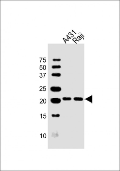
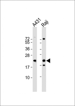
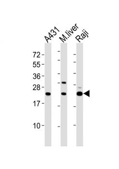
All lanes: Anti-PSMB9 Antibody (C-term) at 1:1000 dilution. Lane 1: A431 whole cell lysates. Lane 2: Raji whole cell lysates. Lysates/proteins at 20 µg per lane. Secondary Goat Anti-Rabbit IgG, (H+L), Peroxidase conjugated at 1/10000 dilution. Predicted band size: 23 kDa. Blocking/Dilution buffer: 5% NFDM/TBST.
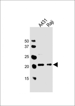
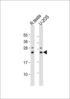
All lanes: Anti-PSMB9 Antibody (C-term) at 1:2000 dilution. Lane 1: A431 whole cell lysates. Lane 2: Raji whole cell lysates. Lysates/proteins at 20 µg per lane. Secondary Goat Anti-Rabbit IgG, (H+L), Peroxidase conjugated at 1/10000 dilution. Predicted band size: 23 kDa. Blocking/Dilution buffer: 5% NFDM/TBST.

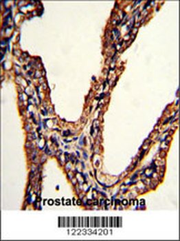
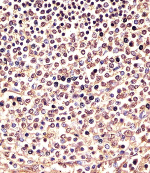
Staining PSMB9 in human spleen sections by Immunohistochemistry (IHC-P - paraformaldehyde-fixed, paraffin-embedded sections). Tissue was fixed with formaldehyde and blocked with 3% BSA for 0.5 hour at room temperature; antigen retrieval was by heat mediation with a citrate buffer (pH6). Samples were incubated with primary antibody (1/25) for 1 hours at 37°C. A undiluted biotinylated goat polyvalent antibody was used as the secondary antibody.
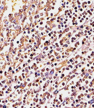
Staining PSMB9 in Human spleen tissue sections by Immunohistochemistry (IHC-P - paraformaldehyde-fixed, paraffin-embedded sections). Tissue was fixed with formaldehyde and blocked with 3% BSA for 0.5 hour at room temperature; antigen retrieval was by heat mediation with a citrate buffer (pH6). Samples were incubated with primary antibody (1/25) for 1 hours at 37°C. A undiluted biotinylated goat polyvalent antibody was used as the secondary antibody.
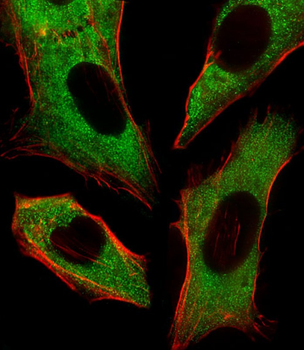
Immunofluorescent analysis of 4% paraformaldehyde-fixed, 0.1% Triton X-100 permeabilized U-2 OS (Human Sarcoma cell line) cells labeling Pdx1 at 1/25 dilution, followed by Alexa Fluor 488-conjugated goat anti-rabbit IgG (1583138) secondary antibody at 1/400 dilution (green). Immunofluorescence image showing cytoplasm staining on U-2 OS cell line. Cytoplasmic actin is detected with Alexa Fluor 555 conjugated with Phalloidin at 1/100 dilution (red).
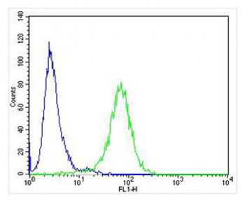
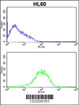
Overlay histogram showing Raji cells stained (green line). The cells were fixed with 4% paraformaldehyde (10 min) and then permeabilized with 90% methanol for 10 min. The cells were then icubated in 2% bovine serum albumin to block non-specific protein-protein interactions followed by the antibody (, 1:25 dilution) for 60 min at 37°C. The secondary antibody used was Alexa Fluor 488 goat anti-rabbit lgG (H+L) (1583138) at 1/400 dilution for 40 min at 37°C. Isotype control antibody (blue line) was rabbit IgG1 (1 μg/1x10^6 cells) used under the same conditions. Acquisition of >10000 events was performed.
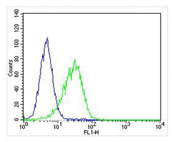
Overlay histogram showing Hela cells stained (green line). The cells were fixed with 4% paraformaldehyde (10 min) and then permeabilized with 90% methanol for 10 min. The cells were then icubated in 2% bovine serum albumin to block non-specific protein-protein interactions followed by the antibody (1:25 dilution) for 60 min at 37°C. The secondary antibody used was Alexa Fluor 488 goat anti-rabbit lgG (H+L) (1583138) at 1/400 dilution for 40 min at 37°C. Isotype control antibody (blue line) was rabbit IgG1 (1 μg/1x10^6 cells) used under the same conditions. Acquisition of >10000 events was performed.
- Item 1 of 4
- Item 1 of 3
PSMB9 Antibody (C-term) [orb1926543]
FC, IHC-P, WB
Human, Mouse
Rabbit
Polyclonal
Unconjugated
100 μl, 50 μl - Item 1 of 1