You have no items in your shopping cart.
Cart summary
Item 1 of 4
Item 1 of 4
PHKG2 Antibody
Catalog Number: orb1263386

| Catalog Number | orb1263386 |
|---|---|
| Category | Antibodies |
| Description | PHKG2 Antibody |
| Target | PHKG2 |
| Clonality | Polyclonal |
| Isotype | Rabbit Ig |
| Conjugation | Unconjugated |
| Reactivity | Human, Mouse |
| Predicted Reactivity | Bovine |
| Form/Appearance | Liquid |
| Concentration | batch dependent |
| Buffer/Preservatives | Supplied in PBS with 0.09% (W/V) sodium azide. |
| Immunogen | This PHKG2 antibody is generated from rabbits immunized with a KLH conjugated synthetic peptide between 41-71 amino acids from the N-terminal region of human PHKG2. |
| UniProt ID | P15735 |
| MW | 46 kDa |
| Tested applications | IHC-P, WB |
| Application notes | For WB starting dilution is: 1:1000For IHC-P starting dilution is: 1:50~100 |
| Antibody Type | Primary Antibody |
| Storage | Maintain refrigerated at 2-8°C for up to 2 weeks. For long term storage store at -20°C in small aliquots to prevent freeze-thaw cycles. |
| Alternative names | Phosphorylase b kinase gamma catalytic chain, live Read more... |
| Note | For research use only |
| NCBI | P15735 |
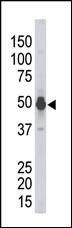
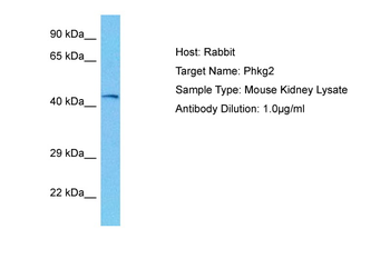
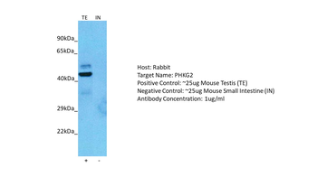
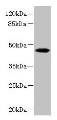
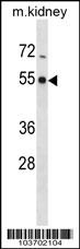
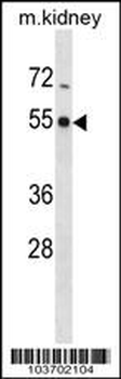
Antibody is used in Western blot to detect PHKG2 in mouse kidney tissue lysate.
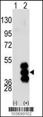
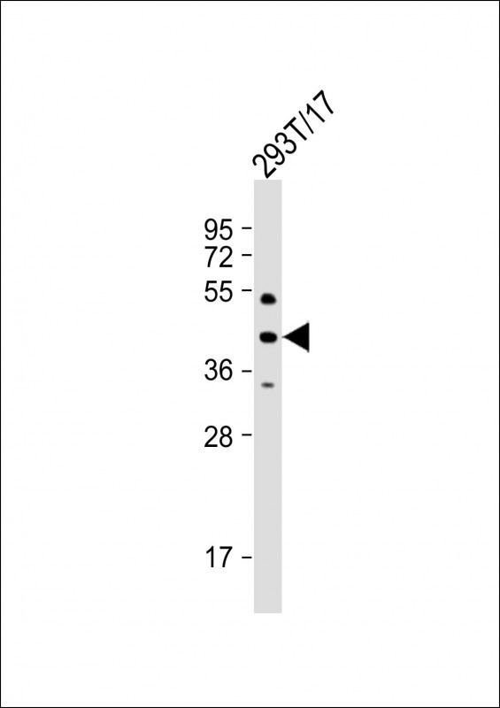
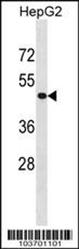
Western blot analysis of PHKG2 using PHKG2 Antibody.293 cell lysates (2 ug/lane) either nontransfected (Lane 1) or transiently transfected with the PHKG2 gene (Lane 2).
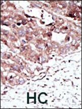
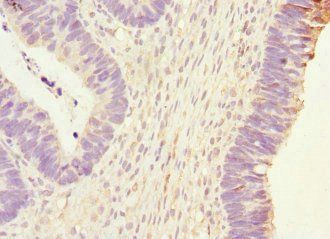
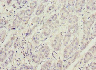
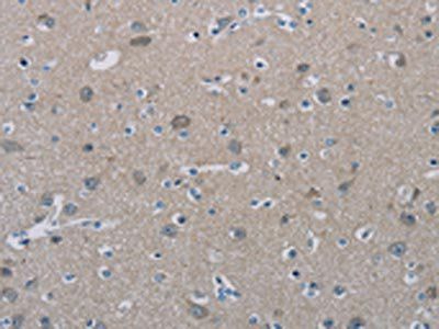
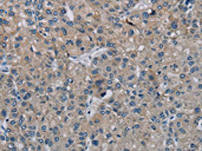
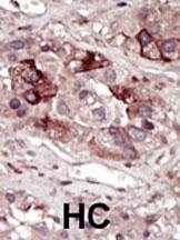
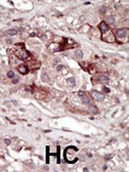
Formalin-fixed and paraffin-embedded human cancer tissue reacted with the primary antibody, which was peroxidase-conjugated to the secondary antibody, followed by AEC staining. BC = breast carcinoma; HC = hepatocarcinoma.
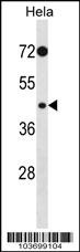
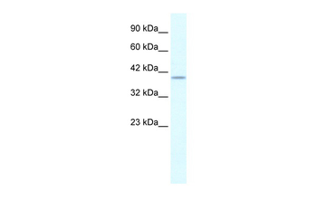
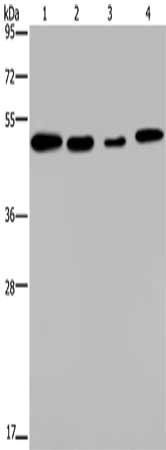
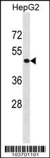
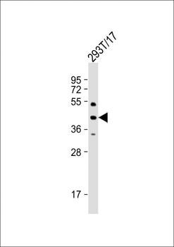
Western blot analysis in Hela cell line lysates (35 ug/lane).
- Item 1 of 4
PHKG2 Rabbit Polyclonal Antibody [orb574500]
IHC, WB
Bovine, Canine, Goat, Guinea pig, Rabbit, Rat, Sheep, Zebrafish
Human, Mouse
Rabbit
Polyclonal
Unconjugated
100 μl - Item 1 of 3
- Item 1 of 3
- Item 1 of 4
- Item 1 of 4
PHKG2 Antibody (Center) [orb1929455]
IHC-P, WB
Human, Mouse
Rabbit
Polyclonal
Unconjugated
100 μl, 50 μl