You have no items in your shopping cart.
Cart summary
Item 1 of 4
Item 1 of 4
PECAM1 Antibody
Catalog Number: orb1252719
| Catalog Number | orb1252719 |
|---|---|
| Category | Antibodies |
| Description | PECAM1 Antibody |
| Species/Host | Mouse |
| Clonality | Monoclonal |
| Clone Number | C31.3 |
| Tested applications | FC, IF, IHC |
| Reactivity | Human |
| Isotype | IgG1, kappa |
| Immunogen | Recombinant human protein |
| Concentration | 0.2 mg/mL |
| Dilution range | Flow Cytometry: 0.5-1 ug/million cellsImmunofluorescence: 1-2 ug/ml Immunohistochemistry (FFPE): 0.5-1 ug/ml for 30 minutes at RT (1)Prediluted format : incubate for 30 min at RT (2)Differences in protocols, secondaries and substrates may require the CD31 antibody to be titered for optimal performance.1. Staining of formalin-fixed tissues requires boiling tissue sections in 1mM EDTA, pH 7.5-8.5, for 10-20 min followed by cooling at RT for 20 minutes.2. The prediluted format is supplied in a dropper bottle and is optimized for use in IHC. After epitope retrieval step (if required), drip mAb solution onto the tissue section and incubate at RT for 30 min. |
| Form/Appearance | Liquid |
| Conjugation | Unconjugated |
| Target | PECAM1 |
| UniProt ID | P16284 |
| Storage | Aliquot and Store at 2-8°C. Avoid freez-thaw cycles. |
| Buffer/Preservatives | PBS with 0.1 mg/ml rAlbumin and 0.05% sodium azide |
| Alternative names | Platelet endothelial cell adhesion molecule, PECAM Read more... |
| Note | For research use only |
| Application notes | Flow Cytometry: 0.5-1 ug/million cellsImmunofluorescence: 1-2 ug/ml Immunohistochemistry (FFPE): 0.5-1 ug/ml for 30 minutes at RT (1)Prediluted format : incubate for 30 min at RT (2)Differences in protocols, secondaries and substrates may require the CD31 antibody to be titered for optimal performance.1. Staining of formalin-fixed tissues requires boiling tissue sections in 1mM EDTA, pH 7.5-8.5, for 10-20 min followed by cooling at RT for 20 minutes.2. The prediluted format is supplied in a dropper bottle and is optimized for use in IHC. After epitope retrieval step (if required), drip mAb solution onto the tissue section and incubate at RT for 30 min. |
| Expiration Date | 12 months from date of receipt. |
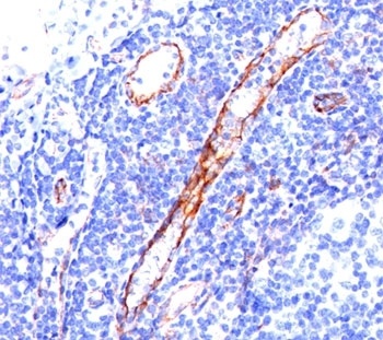
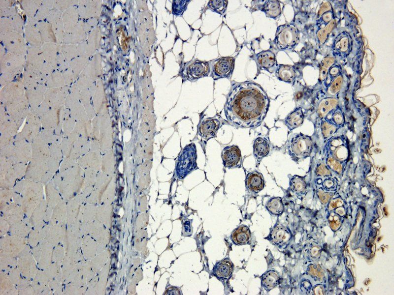
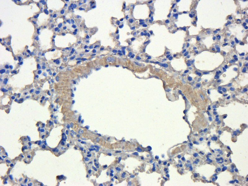
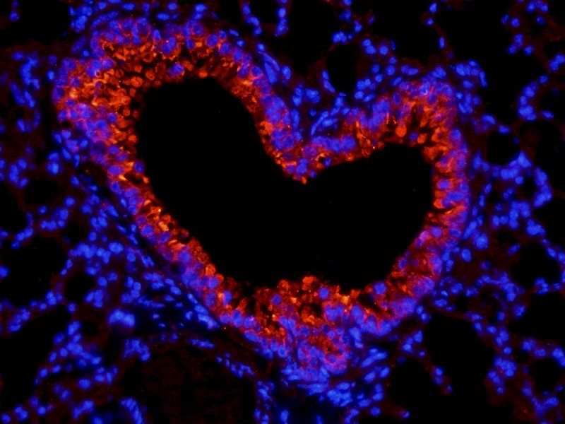
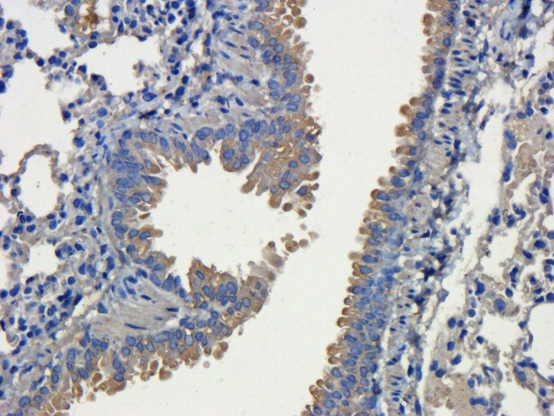
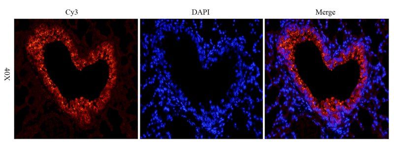
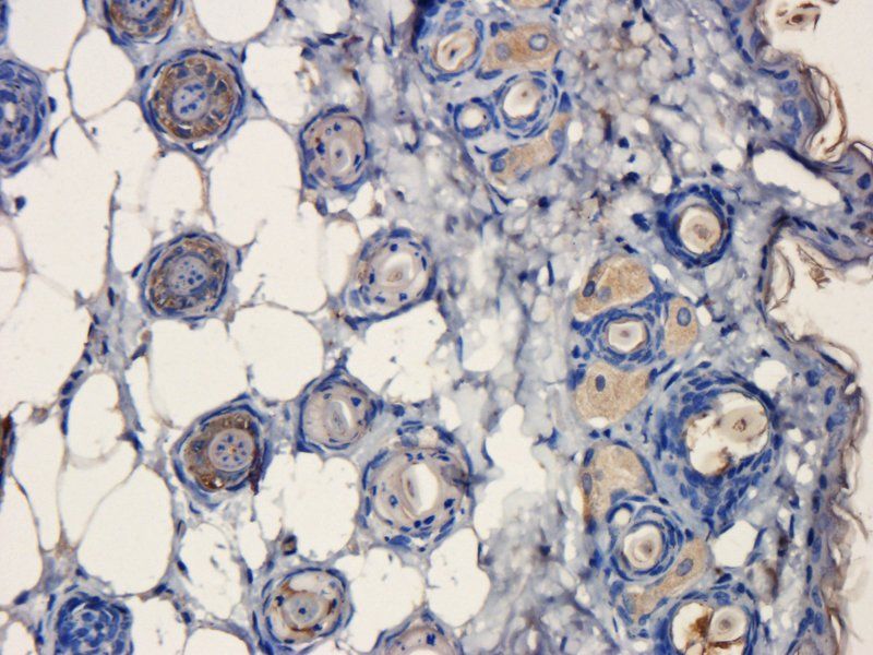
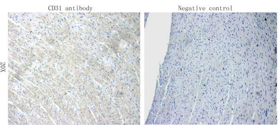
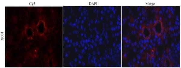
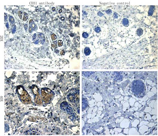
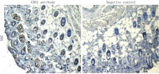
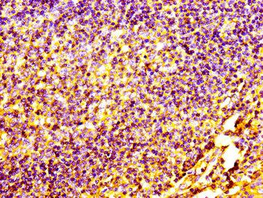
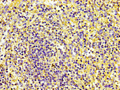
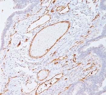
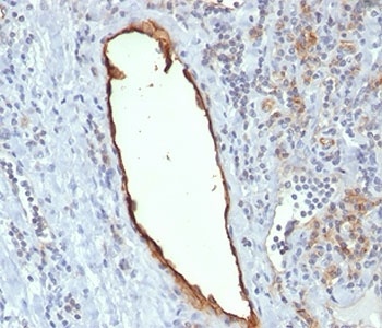
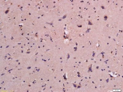
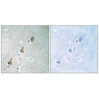
IHC staining of tonsil tissue with CD31 antibody (C31.3).
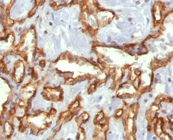
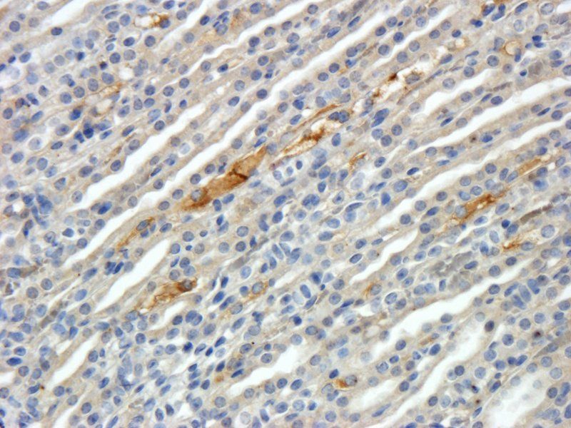
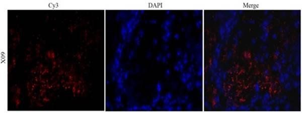
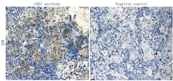
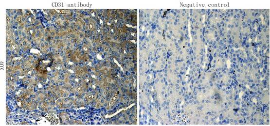
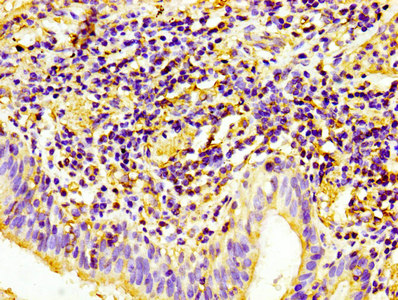
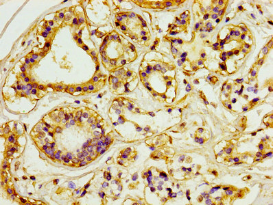
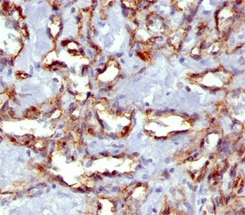
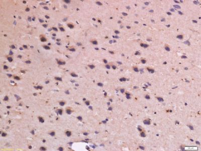
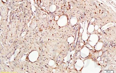
IHC staining of angiocarcinoma with CD31 antibody (C31.3).
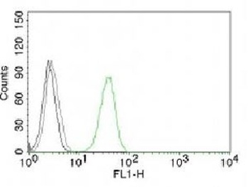
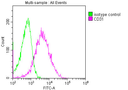
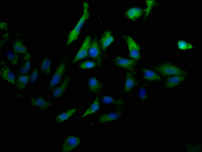
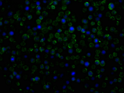
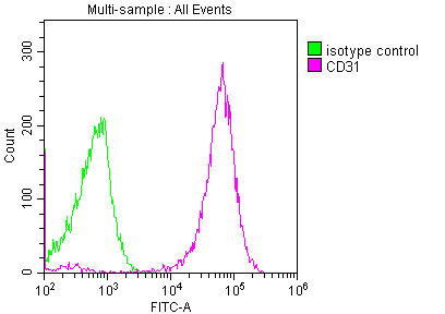
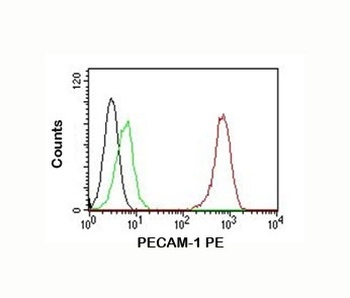
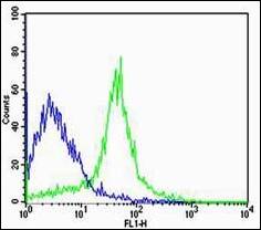
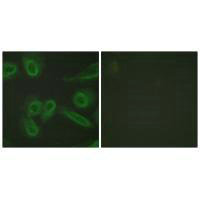
FACS testing of Jurkat cells with Alexa Fluor conjugated CD31 antibody (green) and isotype control (gray).
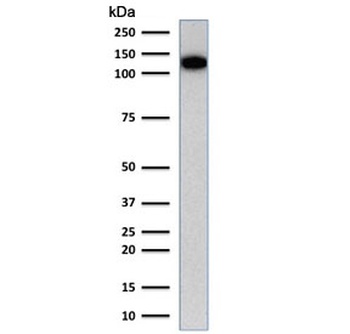
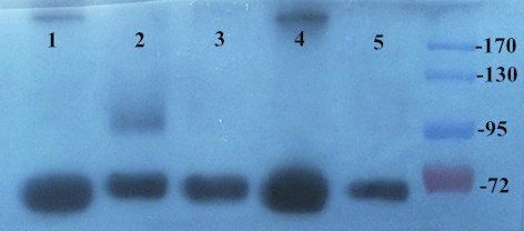
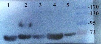
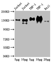
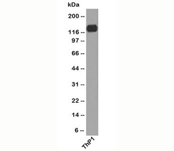
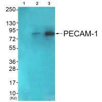
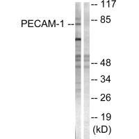
Western blot testing of human Jurkat cell lysate with CD31 antibody (clone C31.1). Expected molecular weight: 83-130 kDa depending on level of glycosylation.
- Item 1 of 20
- Item 1 of 10
- Item 1 of 6
PECAM1 Antibody [orb1252720]
FC, IF, IHC, WB
Human, Primate, Rabbit
Mouse
Monoclonal
Unconjugated
100 μg - Item 1 of 5
CD31 antibody [orb221348]
WB
Equine, Mouse, Rat
Canine, Human, Porcine
Rabbit
Polyclonal
Unconjugated
200 μl, 100 μl, 50 μl - Item 1 of 4
PECAM1 (Ab-713) antibody [orb685571]
ELISA, IF, IHC, WB
Human, Mouse
Rabbit
Polyclonal
Unconjugated
100 μl
Submit a review
Filter by Rating

- 5 stars

- 4 stars
- 3 stars
- 2 stars
- 1 stars