You have no items in your shopping cart.
Cart summary
Item 1 of 4
Item 1 of 4
Pdcd1 Antibody
Catalog Number: orb1671671
| Catalog Number | orb1671671 |
|---|---|
| Category | Antibodies |
| Description | Pdcd1 Antibody |
| Clonality | Recombinant |
| Clone Number | J43 |
| Tested applications | Depletion, ELISA, FC, IHC, IP, NeA |
| Reactivity | Mouse |
| Isotype | IgG lambda |
| Immunogen | This antibody was raised by immunising Armenian hamsters with B12 cells, a PD-1 cDNA transfectant of BHK cells. |
| Concentration | batch dependent |
| Conjugation | Unconjugated |
| Target | Pdcd1 |
| UniProt ID | Q02242 |
| Storage | Store at 4°C for up to 3 months. For longer storage, aliquot and store at -20°C. |
| Buffer/Preservatives | PBS with 0.02% Proclin 300. |
| Alternative names | CD279, PD1, PD 1, Programmed cell death protein 1, Read more... |
| Note | For research use only |
| Expiration Date | 12 months from date of receipt. |
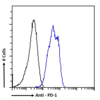
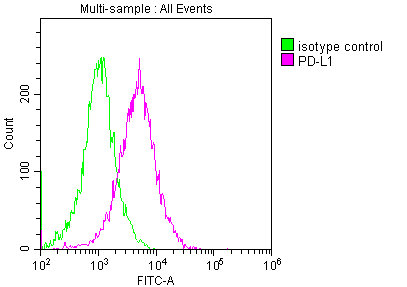
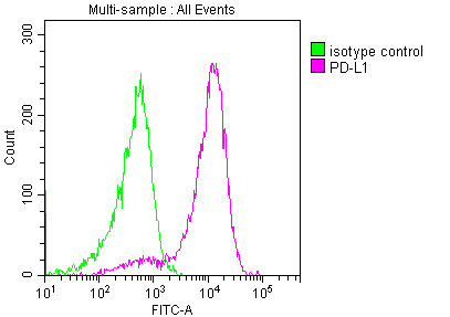
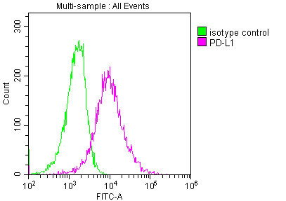
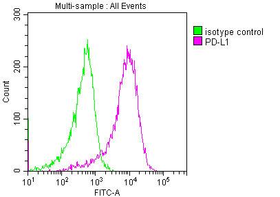
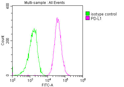
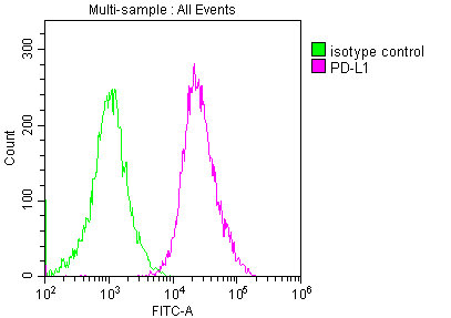
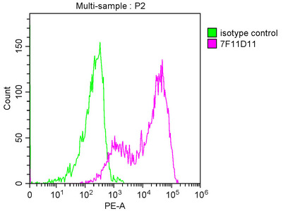
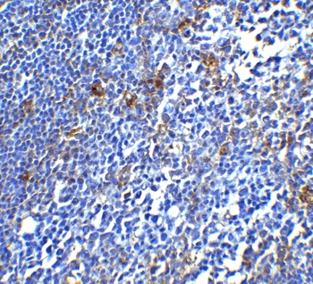
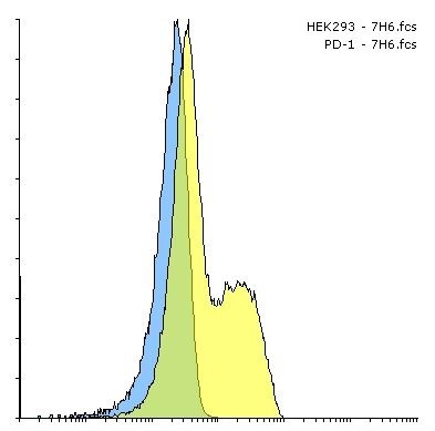
Flow cytometry using the Anti-PD-1 antibody J43. Paraformaldehyde fixed mouse splenocytes permewith 0.5% Triton were stained with anti-unknown specificity antibody (3.0; isotype control - black line) or the r version of J43 (orb1671671 - blue line) at a dilution of 1:100 for 1h at RT. After washing- the bound antibody was detected using a goat anti-r AlexaFluor® 488 antibody at a dilution of 1:1000 and cells analyzed using a FACSCanto flow-cytometer.
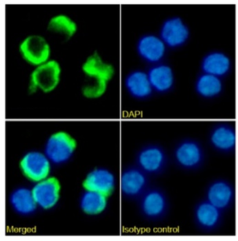
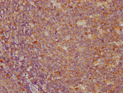
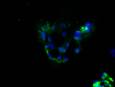
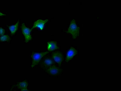
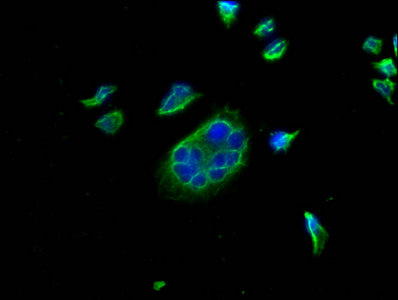
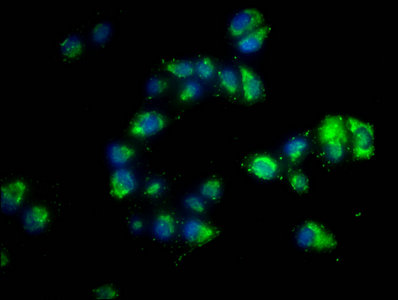
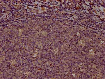
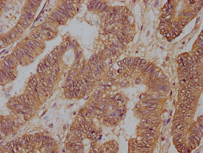
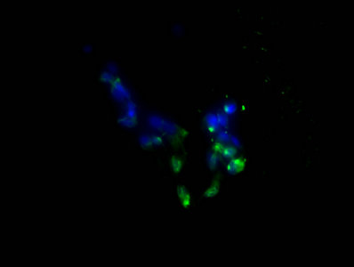
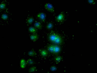
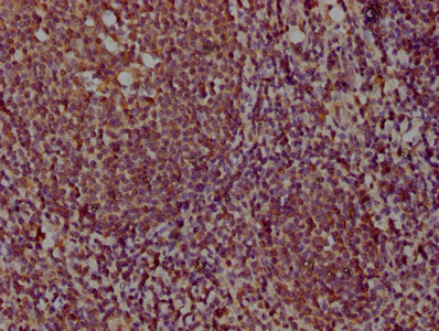
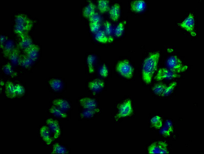
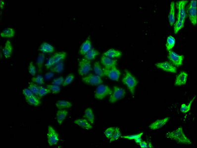
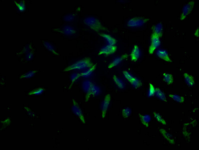
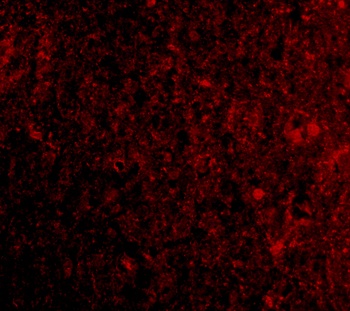
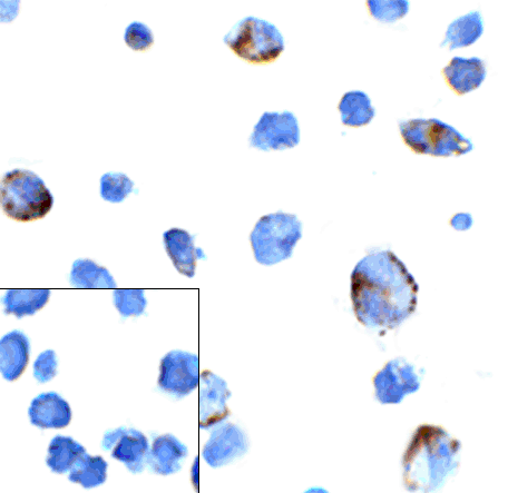
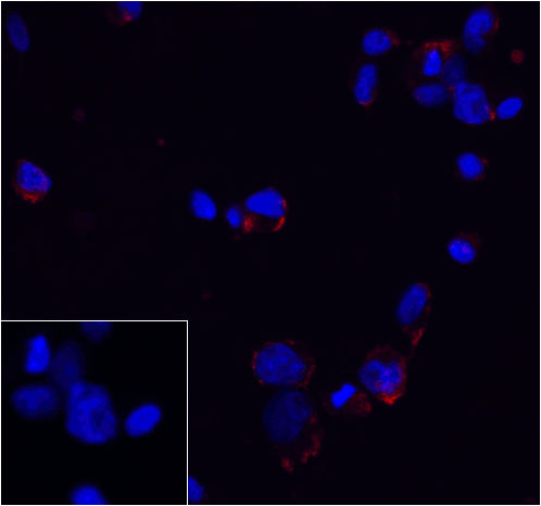
Immunofluorescence staining of mouse splenocytes with anti-PD-1 (J43). Immunofluorescence analysis of paraformaldehyde fixed mouse splenocytes on Shi-fix™ coverslips stained with the chimeric r version of J43 (orb1671671) at 10 ug/ml for 1h followed by Alexa Fluor® 488 secondary antibody (2 ug/ml)- showing membrane staining. The nuclear stain is DAPI (blue). Panels show from left-right- top-bottom orb1671671- DAPI- merged channels and an isotype control. The isotype control was an unknown specificity antibody (3.0) followed by staining with Alexa Fluor® 488 secondary antibody.
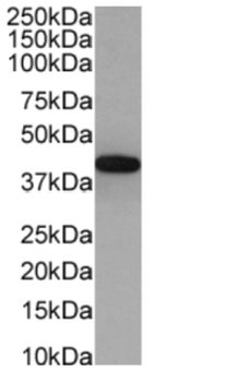
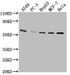
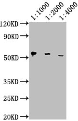
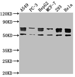
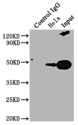
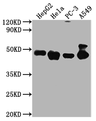
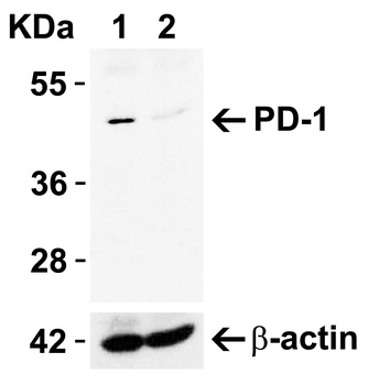
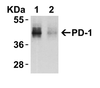
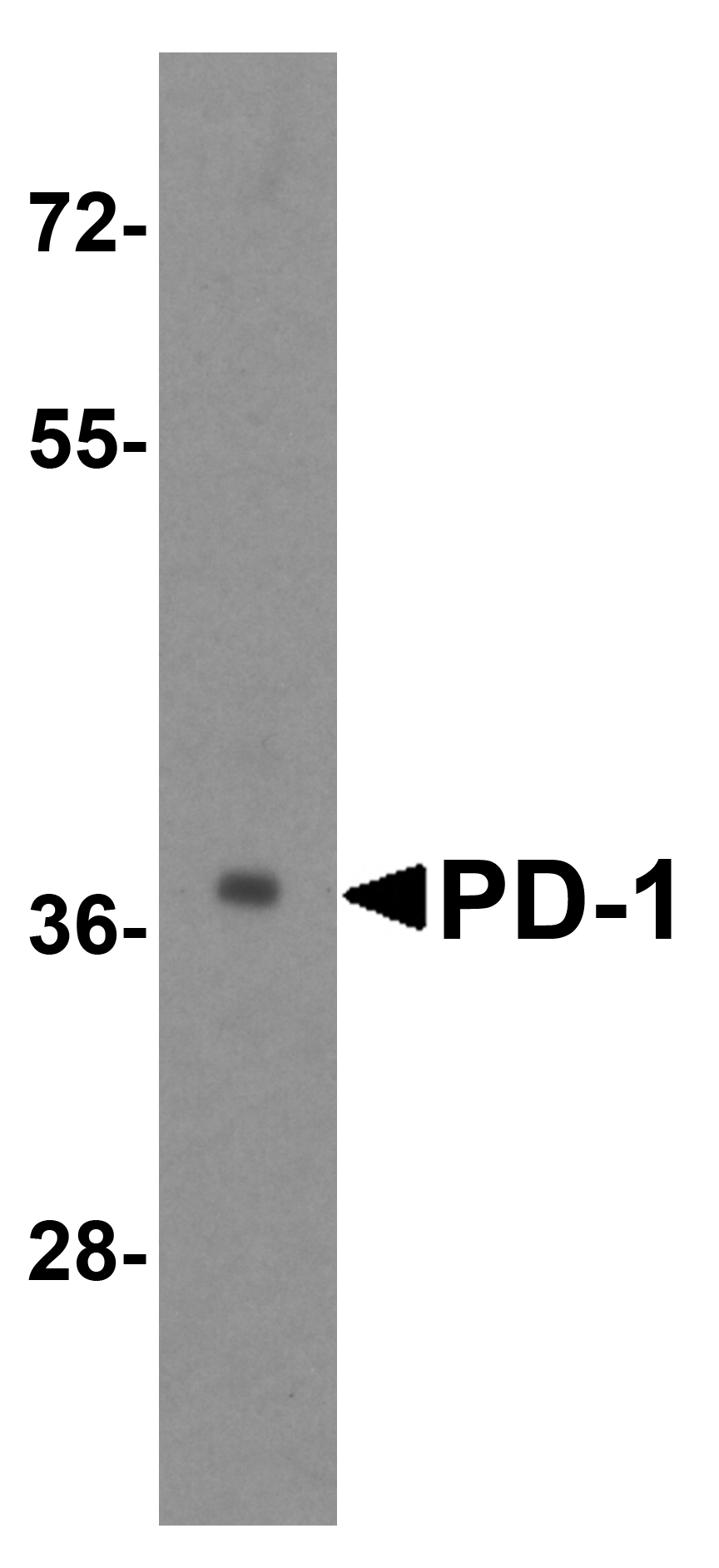
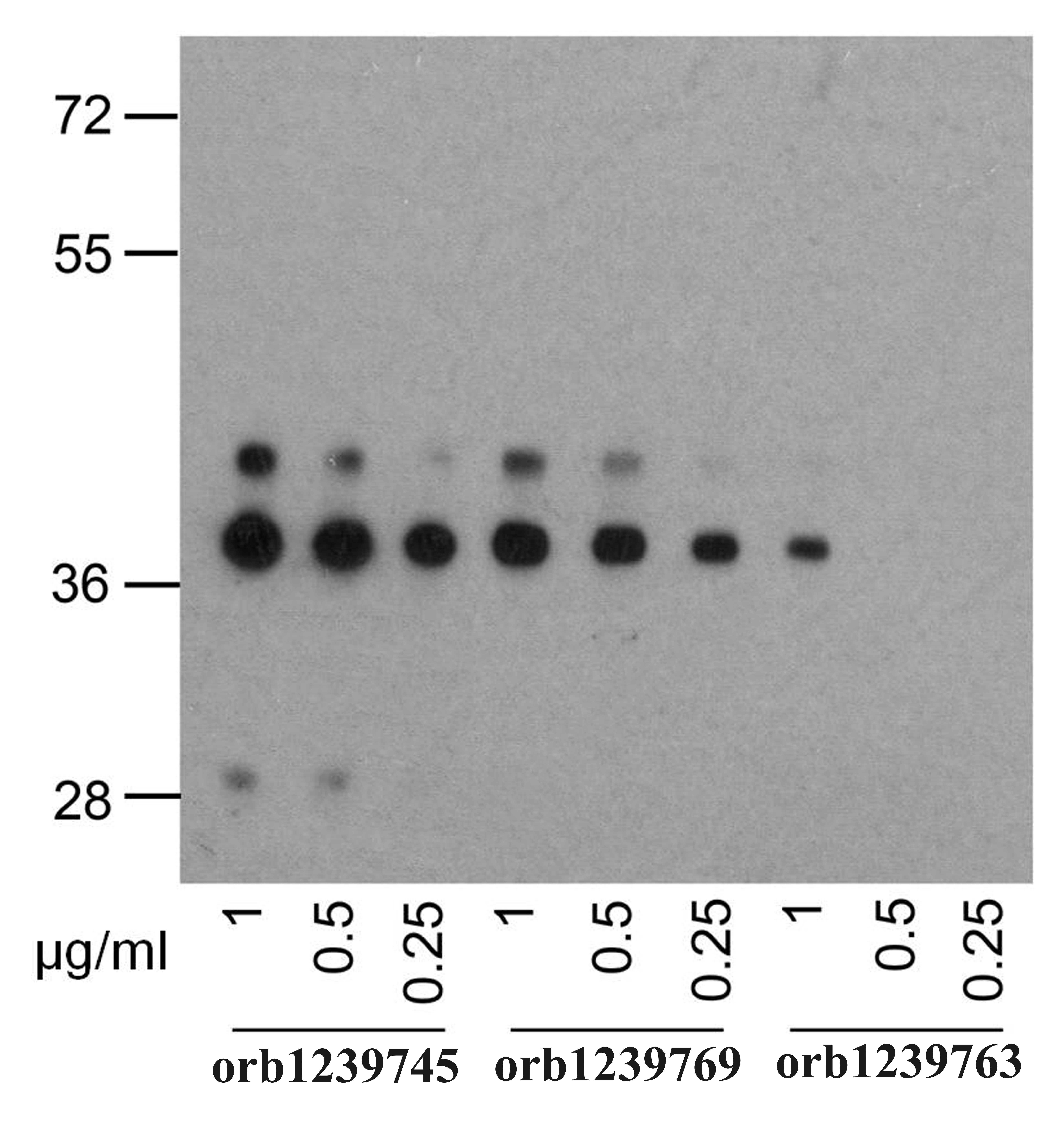
Western Blot using Anti-PD-1 antibody J43. Mouse lymph node tissue lysate (35 ug protein in RIPA buffer) were resolved on a SDS PAGE gel and blots were probed with the chimeric rsion of J43 (orb1671671) at 0.003 ug/ml before detection using an anti-rondary antibody. A primary incubation of 1h was used and protein was detected by chemiluminescence.
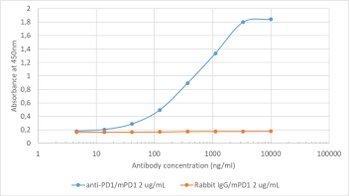
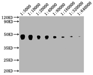
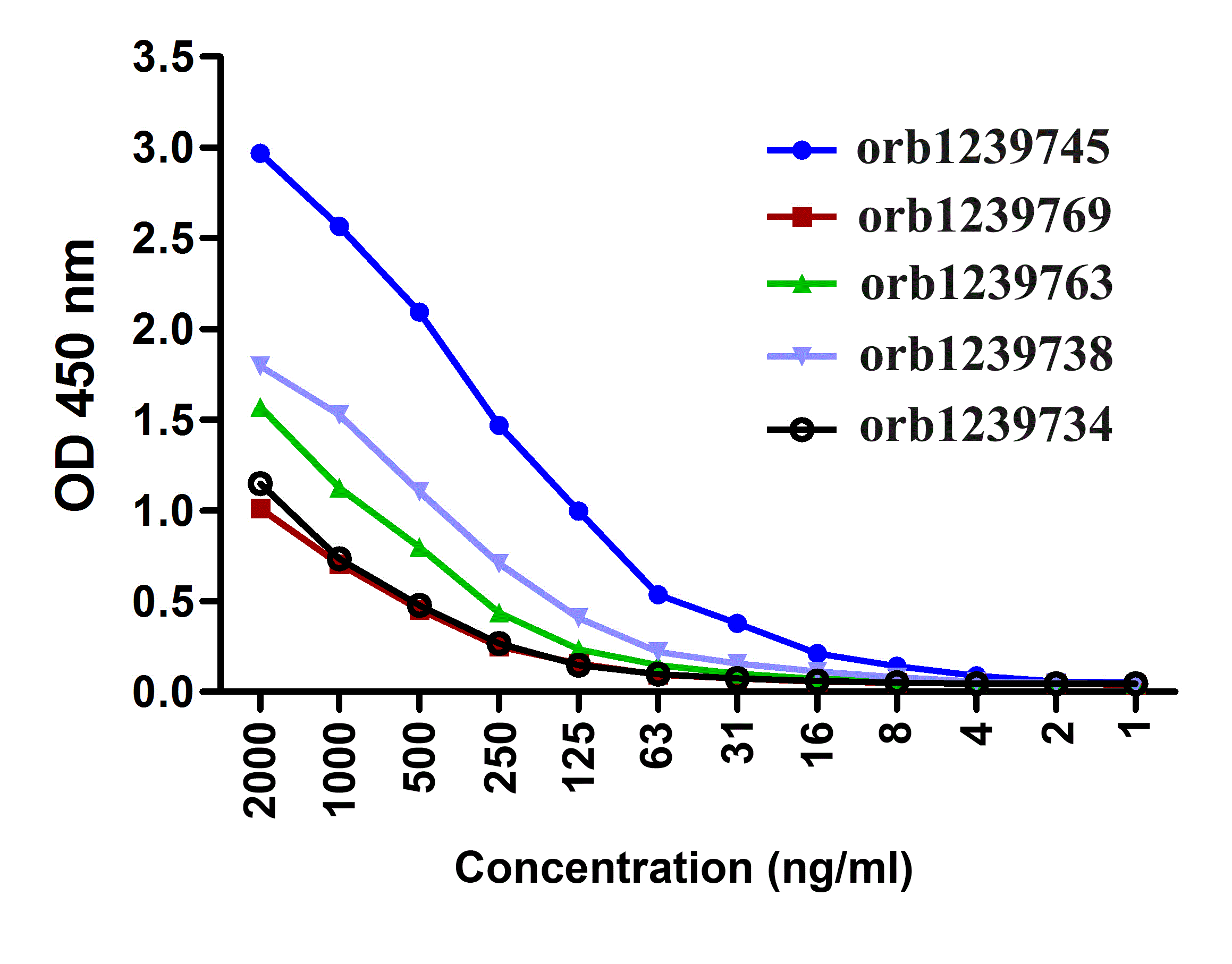
Binding curve of anti-PD-1 antibody J43 (orb1671671) to recombinant mouse PD-1 Fc-Fusion Protein. ELISA Plate coated with recombinant mouse PD-1 Fc-Fusion Protein (Pr00152-1.9; Antibody) at a concentration of 5 ug/ml. A 3-fold serial dilution from 10-000 ng/ml was performed using orb1671671. For detection- a 1:4000 dilution of HRP-lnti-ribody was used.
- Item 1 of 10
- Item 1 of 9
- Item 1 of 9
- Item 1 of 8
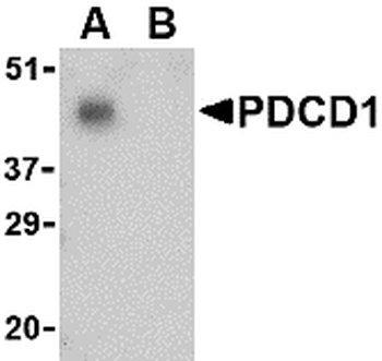
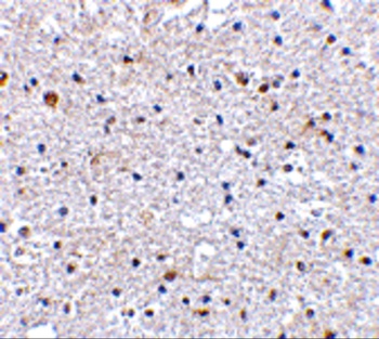
PDCD1 Antibody [orb1239732]
ELISA, IF, IHC-P, WB
Human, Mouse, Rat
Rabbit
Polyclonal
Unconjugated
0.1 mg, 0.02 mg - Item 1 of 8

Submit a review
Filter by Rating

- 5 stars

- 4 stars
- 3 stars
- 2 stars
- 1 stars