You have no items in your shopping cart.
Cart summary
Item 1 of 7
Item 1 of 7
PCSK9 Antibody
Catalog Number: orb1263302

| Catalog Number | orb1263302 |
|---|---|
| Category | Antibodies |
| Description | PCSK9 Antibody |
| Target | PCSK9 |
| Clonality | Polyclonal |
| Isotype | Rabbit Ig |
| Conjugation | Unconjugated |
| Reactivity | Human |
| Form/Appearance | Liquid |
| Concentration | batch dependent |
| Buffer/Preservatives | Supplied in PBS with 0.09% (W/V) sodium azide. |
| Purification | This antibody is purified through a protein A column, followed by peptide affinity purification. |
| Immunogen | This PCSK9 antibody is generated from rabbits immunized with a KLH conjugated synthetic peptide between 144-173 amino acids from the N-terminal region of human PCSK9. |
| UniProt ID | Q8NBP7 |
| MW | 74 kDa |
| Tested applications | FC, IHC-P, WB |
| Application notes | For FACS starting dilution is: 1:25For IHC-P starting dilution is: 1:25For WB starting dilution is: 1:2000 |
| Antibody Type | Primary Antibody |
| Storage | Maintain refrigerated at 2-8°C for up to 2 weeks. For long term storage store at -20°C in small aliquots to prevent freeze-thaw cycles. |
| Alternative names | Proprotein convertase subtilisin/kexin type 9, 342 Read more... |
| Note | For research use only |
| NCBI | Q8NBP7 |
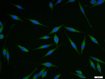
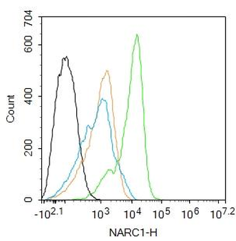
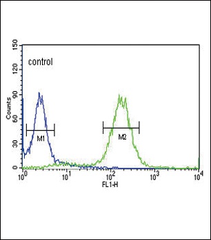
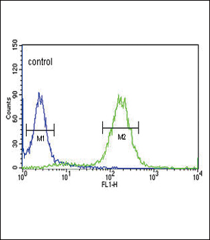
Flow cytometric analysis of HeLa cells using PCSK9 Antibody (green) compared to an isotype control of rabbit IgG (blue). Antibody was diluted at 1:25 dilution. An Alexa Fluor 488 goat anti-rabbit lgG at 1:400 dilution was used as the secondary antibody.
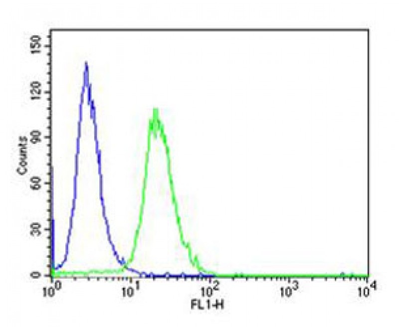
Flow cytometric analysis of HeLa cells using PCSK9 Antibody (green) compared to an isotype control of rabbit IgG (blue). Antibody was diluted at 1:25 dilution. An Alexa Fluor 488 goat anti-rabbit lgG at 1:400 dilution was used as the secondary antibody.

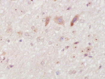
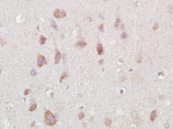
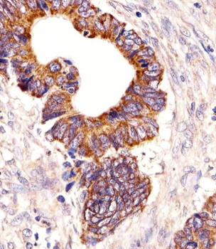
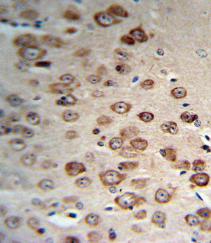
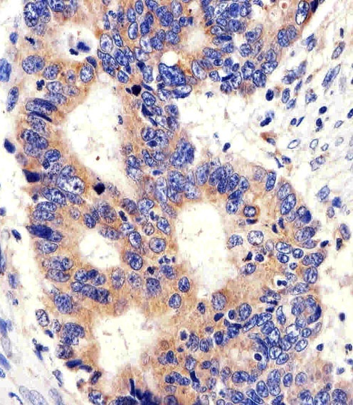
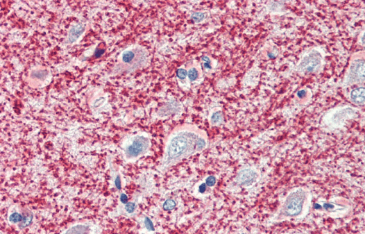
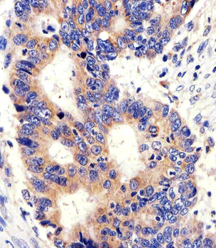
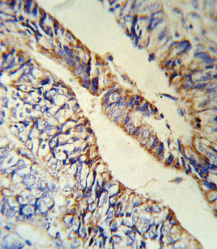
Immunohistochemical analysis of paraffin-embedded H. colorectal carcinoma section using PCSK9 Antibody. Antibody was diluted at 1:25 dilution. A undiluted biotinylated goat polyvalent antibody was used as the secondary, followed by DAB staining.
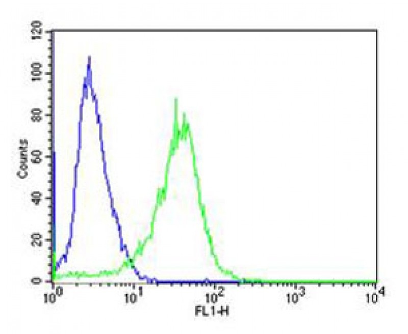
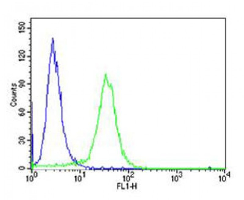
Flow cytometric analysis of A431 cells using PCSK9 Antibody (green) compared to an isotype control of rabbit IgG (blue). Antibody was diluted at 1:25 dilution. An Alexa Fluor 488 goat anti-rabbit lgG at 1:400 dilution was used as the secondary antibody.
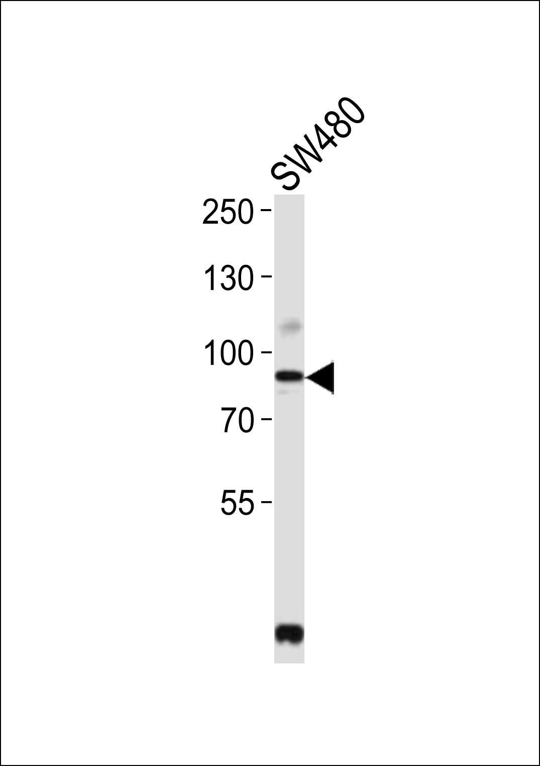
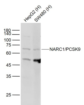
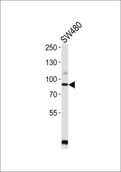
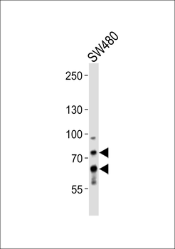
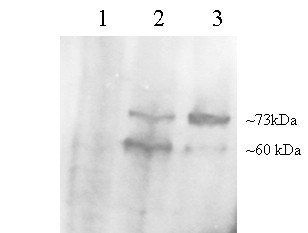
Western blot analysis of lysate from SW480 cell line, using PCSK9 Antibody at 1:2000.
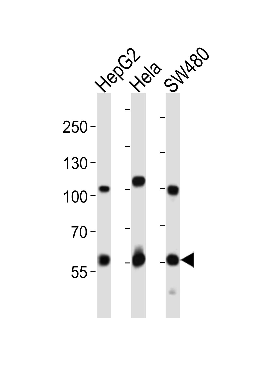
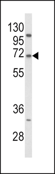
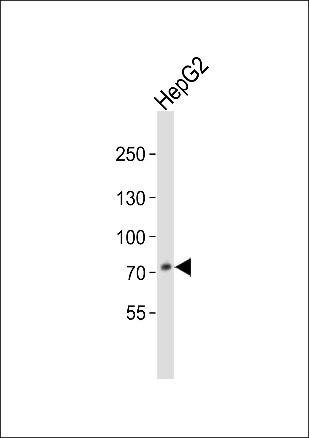
Western blot analysis of lysates from HepG2, Hela, SW480 cell line (from left to right), using PCSK9 Antibody at 1:1000 at each lane.

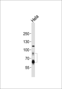
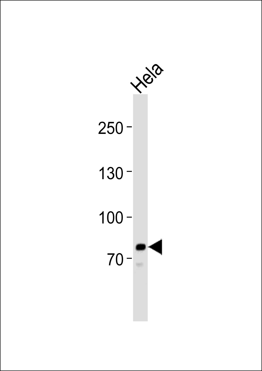
Western blot analysis of lysate from Hela cell line, using PCSK9 Antibody at 1:1000.
- Item 1 of 8
NARC1/PCSK9 Rabbit Polyclonal Antibody [orb100121]
FC, ICC, IF, IHC-Fr, IHC-P, WB
Mouse, Rat
Human, Mouse, Rat
Rabbit
Polyclonal
Unconjugated
100 μl, 50 μl, 200 μl - Item 1 of 7
- Item 1 of 5
- Item 1 of 3
- Item 1 of 5