You have no items in your shopping cart.
Cart summary
Item 1 of 4
Item 1 of 4
PA2G4 Antibody
Catalog Number: orb1264558

| Catalog Number | orb1264558 |
|---|---|
| Category | Antibodies |
| Description | PA2G4 Antibody |
| Target | PA2G4 |
| Clonality | Polyclonal |
| Isotype | Rabbit Ig |
| Conjugation | Unconjugated |
| Reactivity | Human |
| Predicted Reactivity | Mouse, Rat |
| Form/Appearance | Liquid |
| Concentration | batch dependent |
| Buffer/Preservatives | Supplied in PBS with 0.09% (W/V) sodium azide. |
| Purification | This antibody is prepared by Saturated Ammonium Sulfate (SAS) precipitation followed by dialysis |
| Immunogen | This PA2G4 antibody is generated from rabbits immunized with a KLH conjugated synthetic peptide between 228-255 amino acids from the Central region of human PA2G4. |
| UniProt ID | Q9UQ80 |
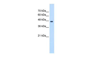
| MW | 44 kDa |
| Tested applications | FC, IF, IHC-P, WB |
| Application notes | For WB starting dilution is: 1:1000For IF starting dilution is: 1:10~50For IHC-P starting dilution is: 1:50~100For FACS starting dilution is: 1:10~50 |
| Antibody Type | Primary Antibody |
| Storage | Maintain refrigerated at 2-8°C for up to 2 weeks. For long term storage store at -20°C in small aliquots to prevent freeze-thaw cycles. |
| Alternative names | Proliferation-associated protein 2G4, Cell cycle p Read more... |
| Note | For research use only |
| NCBI | Q9UQ80 |
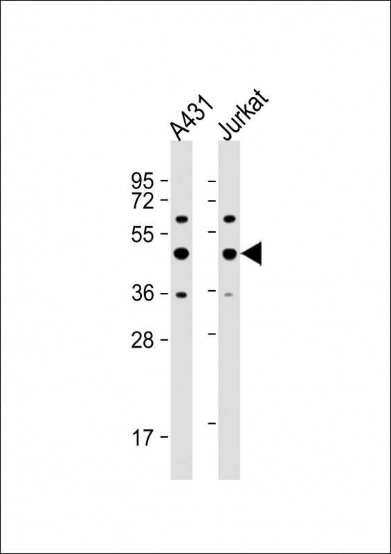
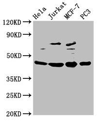
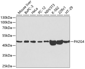
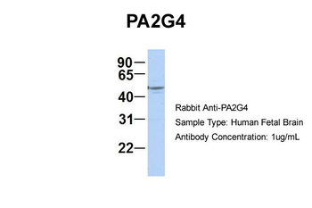
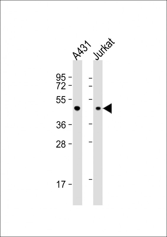
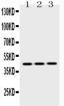
Western Blot at 1:1000 dilution Lane 1: A431 whole cell lysate Lane 2: Jurkat whole cell lysate Lysates/proteins at 20 ug per lane.
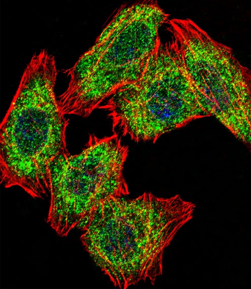
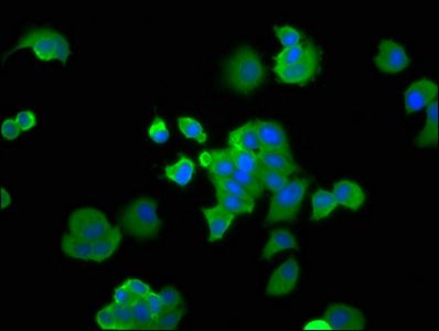
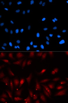
Fluorescent confocal image of U251 cell stained with. U251 cells were fixed with 4% PFA (20 min), permeabilized with Triton X-100 (0.1%, 10 min), then incubated with PA2G4 primary antibody (1:25). For secondary antibody, Alexa Fluor 488 conjugated donkey anti-rabbit antibody (green) was used (1:400). Cytoplasmic actin was counterstained with Alexa Fluor 555 (red) conjugated Phalloidin (7 units/ml). Nuclei were counterstained with DAPI (blue) (10 ug/ml, 10 min).PA2G4 immunoreactivity is localized to Cytoplasm significantly and Nucleus weakly.
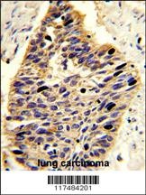
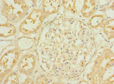
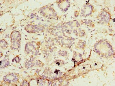
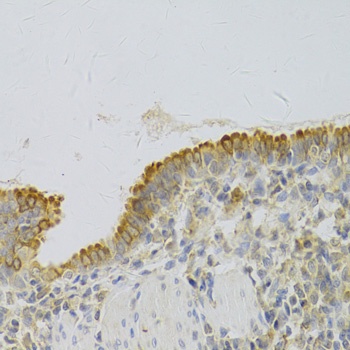
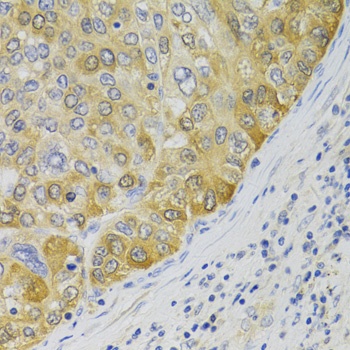
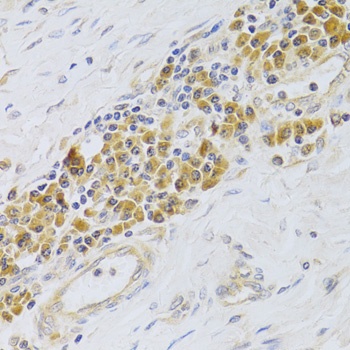
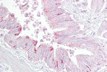
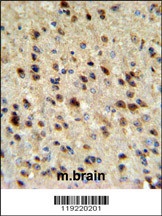
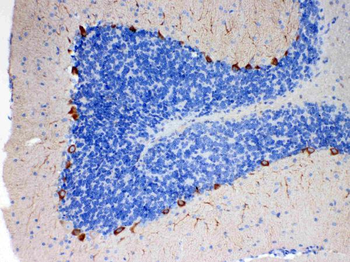
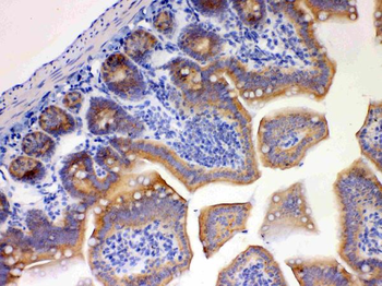
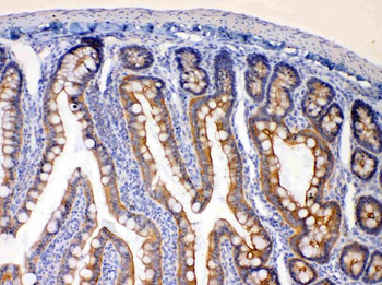
Formalin-fixed and paraffin-embedded human lung carcinoma reacted with, which was peroxidase-conjugated to the secondary antibody, followed by DAB staining.
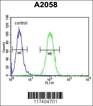
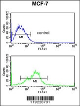
Flow cytometric analysis of A2058 cells (right histogram) compared to a negative control cell (left histogram). FITC-conjugated goat-anti-rabbit secondary antibodies were used for the analysis.
- Item 1 of 4
- Item 1 of 5
- Item 1 of 4
PA2G4 Rabbit Polyclonal Antibody [orb580126]
IHC, WB
Bovine, Canine, Equine, Guinea pig, Mouse, Rabbit, Rat, Zebrafish
Human
Rabbit
Polyclonal
Unconjugated
100 μl - Item 1 of 4
- Item 1 of 4
Anti-EBP1/PA2G4 Antibody [orb389428]
IHC, WB
Human, Mouse, Rat
Rabbit
Polyclonal
Unconjugated
10 μg, 100 μg