You have no items in your shopping cart.
Cart summary
Item 1 of 12
Item 1 of 12
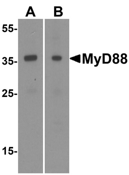
MyD88 Antibody
Catalog Number: orb215902
| Catalog Number | orb215902 |
|---|---|
| Category | Antibodies |
| Description | MyD88 Antibody |
| Species/Host | Rabbit |
| Clonality | Polyclonal |
| Tested applications | ELISA, FC, ICC, IF, IHC, IHC-Fr, WB |
| Reactivity | Human, Mouse, Rat |
| Isotype | Rabbit IgG |
| Immunogen | E.coli-derived human MyD88 recombinant protein (Position: A44-F264). Human MyD88 shares 84% and 83% amino acid (aa) sequences identity with mouse and rat MyD88, respectively. |
| Concentration | Adding 0.2 ml of distilled water will yield a concentration of 500 μg/ml. |
| Dilution range | Western blot, 0.1-0.5μg/ml Immunohistochemistry (Paraffin-embedded Section), 0.5-1μg/ml Immunohistochemistry (Frozen Section), 0.5-1μg/ml Immunocytochemistry, 0.5-1μg/ml Immunofluorescence, 2μg/ml Flow Cytometry, 1-3μg/1x106 cells ELISA (Cap), 1-5μg/ml |
| Form/Appearance | Lyophilized |
| Conjugation | Unconjugated |
| MW | 33233 MW |
| UniProt ID | P35354 |
| Storage | Store at -20˚C for one year from date of receipt. After reconstitution, at 4˚C for one month. It can also be aliquotted and stored frozen at -20˚C for six months. Avoid repeated freeze-thaw cycles. |
| Alternative names | Myeloid differentiation primary response protein M Read more... |
| Note | For research use only |
| Application notes | WB: The detection limit for MyD88 is approximately 0.25ng/lane under reducing conditions. Tested Species: In-house tested species with positive results. By Heat: Boiling the paraffin sections in 10mM citrate buffer, pH6.0, for 20mins is required for the staining of formalin/paraffin sections. Other applications have not been tested. Optimal dilutions should be determined by end users. . Add 0.2ml of distilled water will yield a concentration of 500ug/ml. |
| Expiration Date | 12 months from date of receipt. |
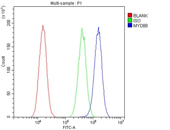
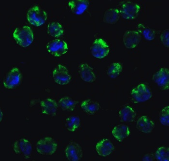
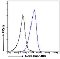
Flow Cytometry analysis of A549 cells using anti-MYD88 antibody (Blue line).Isotype control antibody (Green line) was rabbit IgG .Unlabelled sample (Red line) was also used as a control.
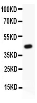
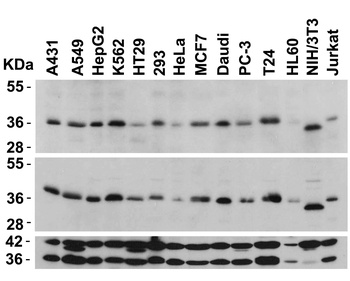
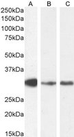
WB analysis of MYD88 using anti-MYD88 antibody.Lane 1:Recombinant Human MYD88 Protein 0.5ng.
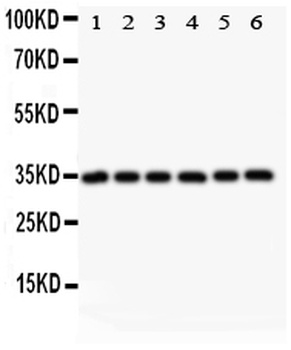
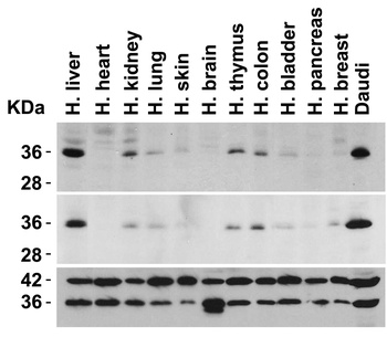
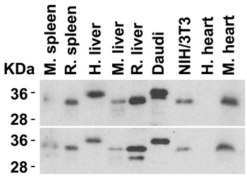
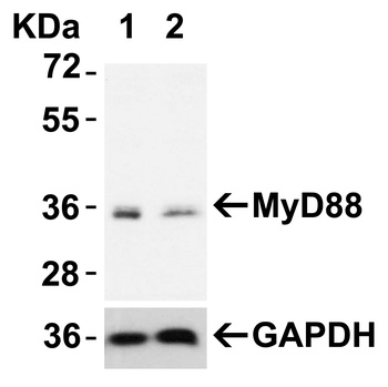
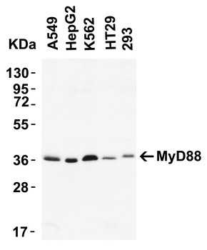
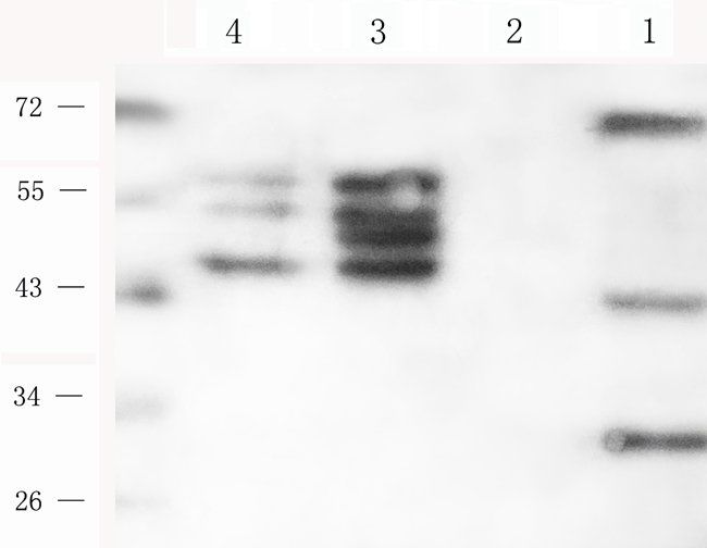
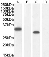
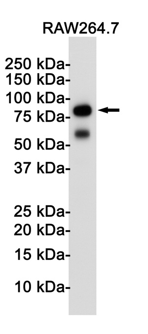
WB analysis of MYD88 using anti-MYD88 antibody.Lane 1:Rat Cardiac Muscle tissue;2:HELA cell;3:MCF cell;4:HEPG2 cell;5:JURKAT cell;6:RAJI Cell.
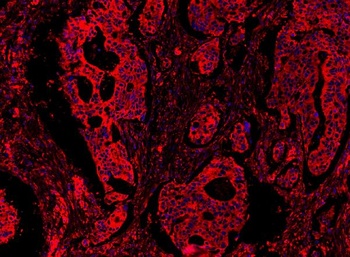
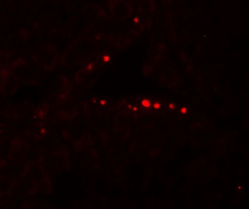
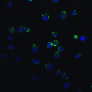
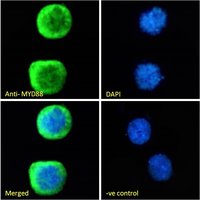
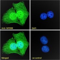
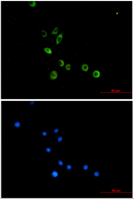
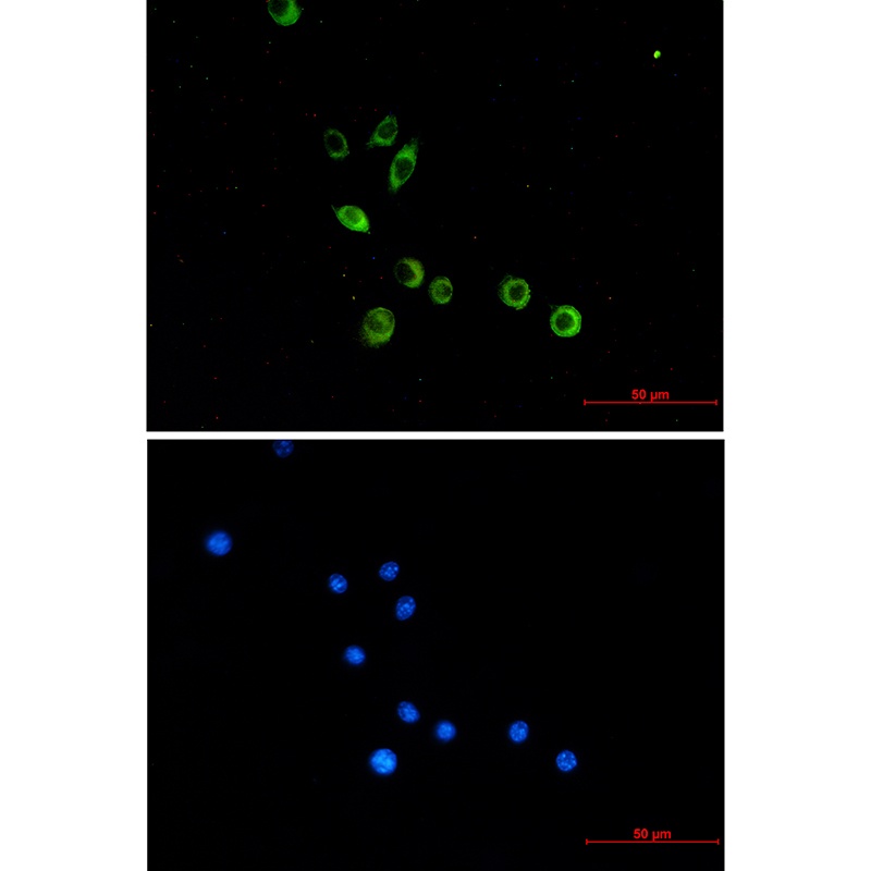
IF analysis of MYD88 using anti-MYD88 antibody.MYD88 was detected in paraffin-embedded section of human colon cancar tissues.
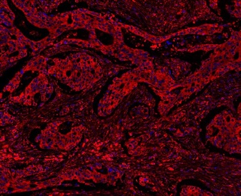
IF analysis of MYD88 using anti-MYD88 antibody.MYD88 was detected in paraffin-embedded section of human colon cancar tissues.
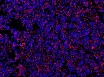
IF analysis of MYD88 using anti-MYD88 antibody.MYD88 was detected in paraffin-embedded section of human tonsil tissues.
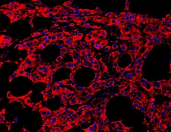
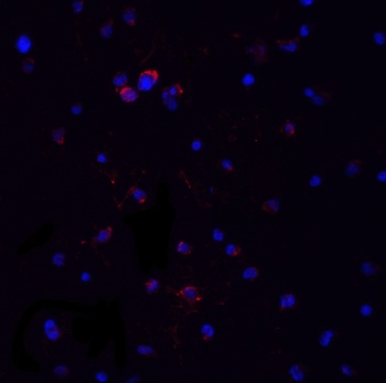
IF analysis of MYD88 using anti-MYD88 antibody.MYD88 was detected in paraffin-embedded section of human mammary cancar tissues.
IF analysis of MYD88 using anti-MYD88 antibody.MYD88 was detected in paraffin-embedded section of human mammary cancar tissues.
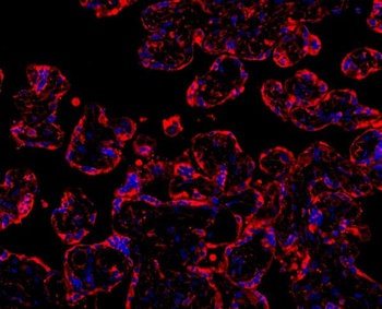
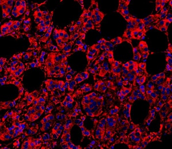
IF analysis of MYD88 using anti-MYD88 antibody.MYD88 was detected in paraffin-embedded section of human placenta tissues.
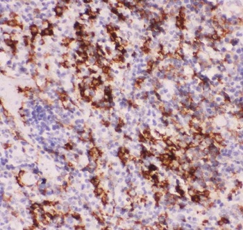
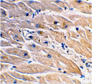
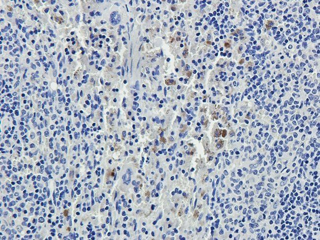
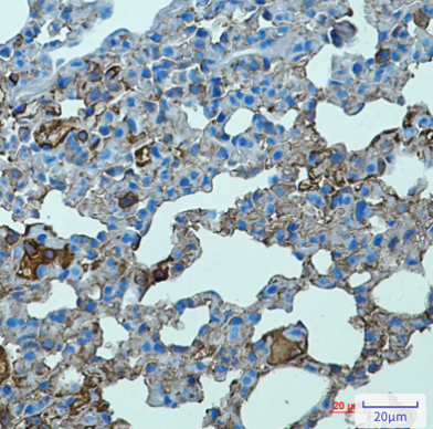
IHC analysis of MYD88 using anti-MYD88 antibody.MYD88 was detected in paraffin-embedded section of rat lung tissue.
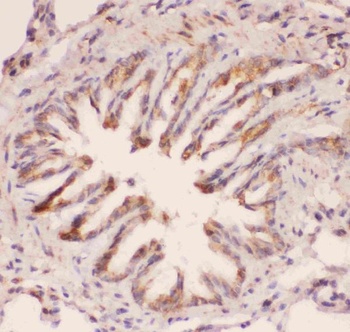
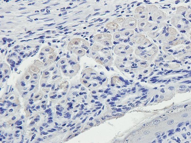
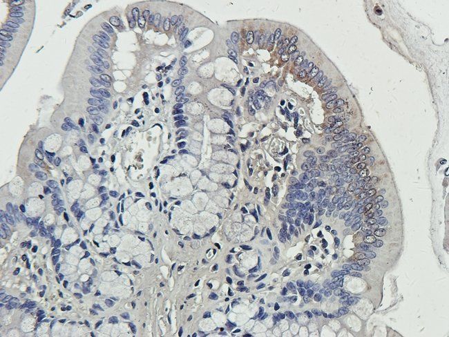
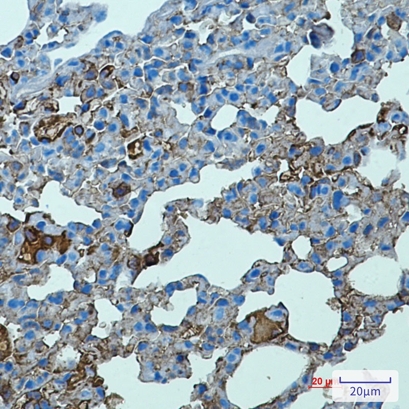
IHC analysis of MYD88 using anti-MYD88 antibody.MYD88 was detected in paraffin-embedded section of human intestinal cancer tissue.
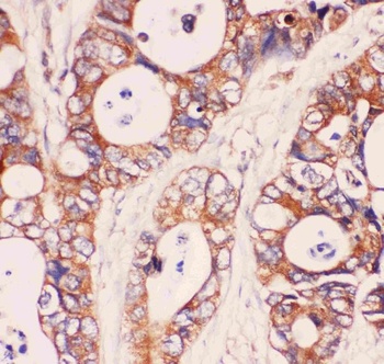
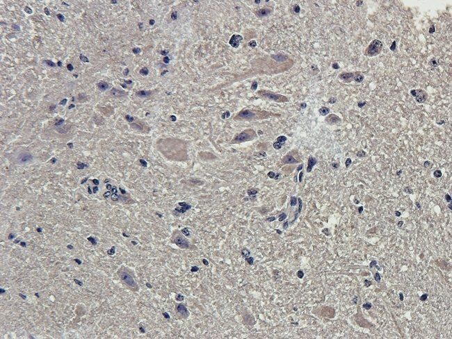
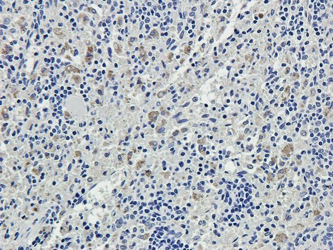
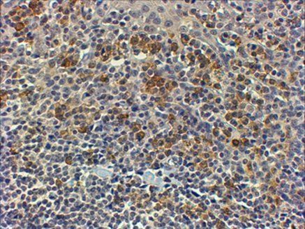
IHC analysis of MYD88 using anti-MYD88 antibody.MYD88 was detected in paraffin-embedded section of mouse spleen tissue.
- Item 1 of 13
MYD88 Antibody [orb1239647]
ELISA, IF, IHC-P, IP, WB
Bovine, Gallus, Porcine, Sheep
Human, Mouse, Rat
Rabbit
Polyclonal
Unconjugated
0.1 mg, 0.02 mg - Item 1 of 11
MYD88 Antibody [orb1239635]
ELISA, IF, IP, WB
Bovine, Gallus, Porcine, Sheep
Human, Mouse, Rat
Rabbit
Polyclonal
Unconjugated
0.1 mg, 0.02 mg - Item 1 of 6
MYD88 antibody [orb420074]
IHC-P, WB
Guinea pig, Human, Mouse, Rat
Rabbit
Polyclonal
Unconjugated
100 μg, 200 μg - Item 1 of 6
MYD88 antibody [orb18963]
ELISA, FC, IF, IHC, WB
Canine, Human, Mouse, Rat
Goat
Polyclonal
Unconjugated
100 μg - Item 1 of 6
Tlr2 Antibody [orb1565339]
ICC, IHC-Fr, IHC-P, WB
Mouse
Rabbit
Monoclonal
Unconjugated
100 μl, 50 μl, 20 μl


Submit a review
Filter by Rating

- 5 stars

- 4 stars
- 3 stars
- 2 stars
- 1 stars