You have no items in your shopping cart.
Cart summary
Item 1 of 5
Item 1 of 5
MSX1 Antibody
Catalog Number: orb1265639

| Catalog Number | orb1265639 |
|---|---|
| Category | Antibodies |
| Description | MSX1 Antibody |
| Species/Host | Rabbit |
| Clonality | Polyclonal |
| Tested applications | IF, WB |
| Predicted Reactivity | Bovine |
| Reactivity | Human, Mouse |
| Isotype | Rabbit Ig |
| Immunogen | This MSX1 antibody is generated from rabbits immunized with a KLH conjugated synthetic peptide between 111-138 amino acids from the Central region of human MSX1. |
| Antibody Type | Primary Antibody |
| Concentration | batch dependent |
| Form/Appearance | Liquid |
| Conjugation | Unconjugated |
| MW | 31 kDa |
| Target | MSX1 |
| UniProt ID | P28360 |
| NCBI | P28360 |
| Storage | Maintain refrigerated at 2-8°C for up to 2 weeks. For long term storage store at -20°C in small aliquots to prevent freeze-thaw cycles. |
| Buffer/Preservatives | Supplied in PBS with 0.09% (W/V) sodium azide. |
| Alternative names | Homeobox protein MSX-1, Homeobox protein Hox-7, Ms Read more... |
| Note | For research use only |
| Application notes | For WB starting dilution is: 1:2000For IF starting dilution is: 1:10~50 |
| Expiration Date | 12 months from date of receipt. |
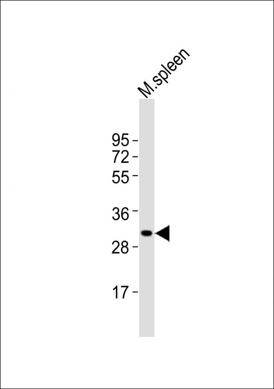
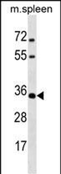
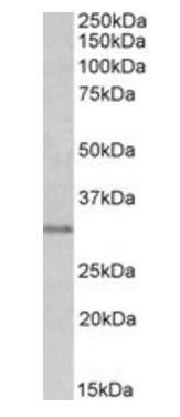
Western Blot at 1:2000 dilution + mouse spleen lysates Lysates/proteins at 20 ug per lane.
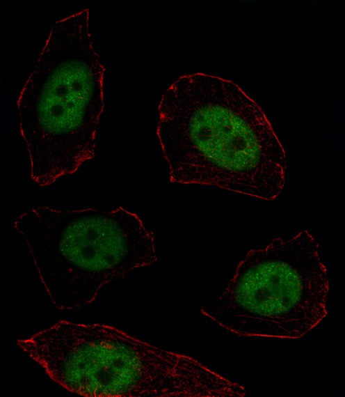
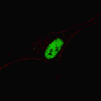
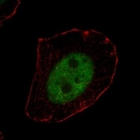
Fluorescent image of U251 cell stained with MSX1 Antibody. U251 cells were fixed with 4% PFA (20 min), permeabilized with Triton X-100 (0.1%, 10 min), then incubated with MSX1 primary antibody (1:25). For secondary antibody, Alexa Fluor 488 conjugated donkey anti-rabbit antibody (green) was used (1:400). Cytoplasmic actin was counterstained with Alexa Fluor 555 (red) conjugated Phalloidin (7 units/ml).MSX1 immunoreactivity is localized to Nucleus significantly.
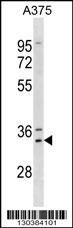
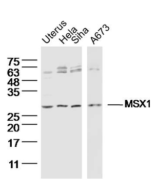
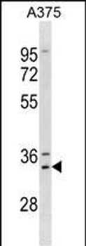
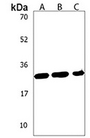
Western blot analysis in A375 cell line lysates (35 ug/lane).
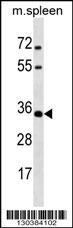
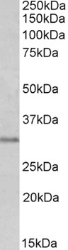
Western blot analysis in mouse spleen tissue lysates (35 ug/lane).

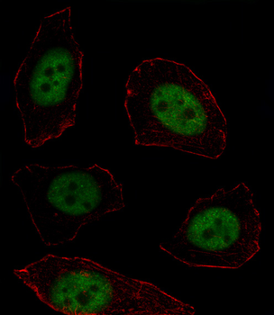
Fluorescent confocal image of SY5Y cells stained with MSX1 antibody. SY5Y cells were fixed with 4% PFA (20 min), permeabilized with Triton X-100 (0.2%, 30 min). Cells were then incubated with MSX1 primary antibody (1:200, 2 h at room temperature). For secondary antibody, Alexa Fluor 488 conjugated donkey anti-rabbit antibody (green) was used (1:1000, 1h). Nuclei were counterstained with Hoechst 33342 (blue) (10 ug/ml, 5 min). Note the highly specific localization of the MSX1 mainly to the nucleus.
- Item 1 of 3
MSX1 Rabbit Polyclonal Antibody [orb2059]
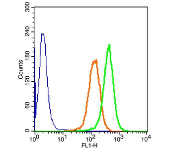
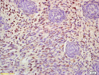
FC, IF, IHC-Fr, IHC-P, WB
Bovine, Canine, Rat
Human, Mouse
Rabbit
Polyclonal
Unconjugated
50 μl, 100 μl, 200 μl - Item 1 of 5
- Item 1 of 2
Goat anti-MSX1 Antibody [orb18823]
ELISA, IHC, WB
Bovine, Human, Mouse, Porcine, Rat
Goat
Polyclonal
Unconjugated
100 μg - Item 1 of 1
MSX1 Antibody [orb1537784]
ELISA, IHC, IHC-P
Bovine, Human, Monkey, Mouse, Porcine, Rat
Goat
Polyclonal
Unconjugated
50 μg - Item 1 of 2