You have no items in your shopping cart.
Cart summary
Item 1 of 5
Item 1 of 5
MART-1 / Melan-A Antibody
Catalog Number: orb2637709

| Catalog Number | orb2637709 |
|---|---|
| Category | Antibodies |
| Description | This antibody recognizes a protein doublet of 20-22kDa, identified as MART-1 (Melanoma Antigen Recognized by T cells 1) or Melan-A. MART-1 is a melanocyte differentiation antigen recognized by autologous cytotoxic T lymphocytes. There are seven other melanoma associated antigens recognized by autologous cytotoxic T cells: MAGE-1, MAGE-3, tyrosinase, gp100, gp75, BAGE-1, and GAGE-1. Subcellular fractionation shows that MART-1 is present in melanosomes and endoplasmic reticulum. This MART-1 antibody labels melanomas and other tumors showing melanocytic differentiation. It is also a useful positive-marker for angiomyolipomas. The antibody does not stain tumor cells of epithelial, lymphoid, glial, or mesenchymal origin. |
| Species/Host | Mouse |
| Clonality | Monoclonal |
| Clone Number | M2-7C10 |
| Tested applications | IHC-P, WB |
| Reactivity | Human |
| Isotype | Mouse IgG2b, lambda |
| Immunogen | Recombinant human MART-1 protein was used as the immunogen for this antibody. |
| Antibody Type | Primary Antibody |
| Dilution range | Western blot: 2-4ug/ml,Immunohistochemistry (FFPE): 1-2ug/ml for 30 min at RT,Prediluted IHC only format: incubate for 30 min at RT (1) |
| Purity | Protein G affinity chromatography |
| Conjugation | Unconjugated |
| Formula | 0.2 mg/ml in 1X PBS with 0.1 mg/ml BSA (US sourced) and 0.05% sodium azide |
| Hazard Information | This MART-1 antibody is available for research use only. |
| Entrez | 2315 |
| Storage | Maintain refrigerated at 2-8°C for up to 2 weeks. For long term storage store at -20°C in small aliquots to prevent freeze-thaw cycles. |
| Buffer/Preservatives | 0.2 mg/ml in 1X PBS with 0.1 mg/ml rAlbumin (US sourced) and 0.05% sodium azide |
| Note | For research use only |
| Application notes | The concentration stated for each application is a general starting point. Variations in protocols, secondaries and substrates may require the MART-1 antibody to be titered up or down for optimal performance.1. The prediluted format is supplied in a dropper bottle and is optimized for use in IHC. After epitope retrieval step (if required), drip mAb solution onto the tissue section and incubate at RT for 30 min. |
| Expiration Date | 12 months from date of receipt. |

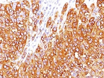
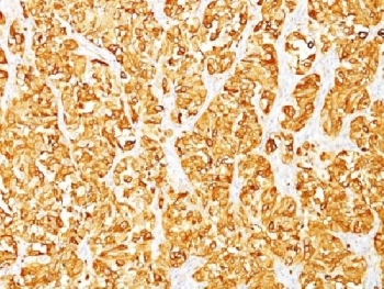
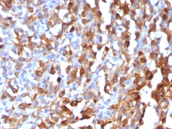
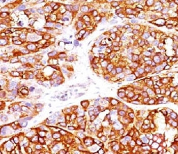
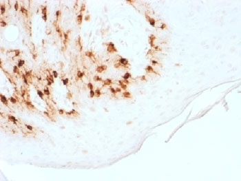
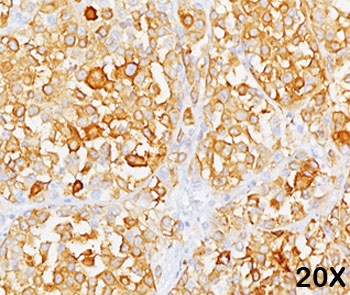
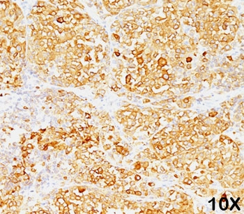
IHC testing of FFPE human melanoma stained with MART-1 antibody (clone M2-7C10). Note cytoplasmic staining of cells. HIER: boil tissue sections in pH9 10mM Tris with 1mM EDTA for 10-20 min.

IHC testing of FFPE human melanoma stained with MART-1 antibody (clone M2-7C10). Note cytoplasmic staining of cells. HIER: boil tissue sections in pH9 10mM Tris with 1mM EDTA for 10-20 min.

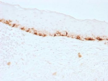
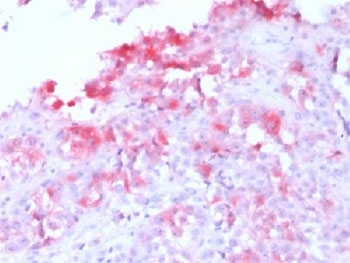
IHC testing of FFPE human skin stained with MART-1 antibody (clone M2-7C10). Note cytoplasmic staining of cells. HIER: boil tissue sections in pH9 10mM Tris with 1mM EDTA for 10-20 min.
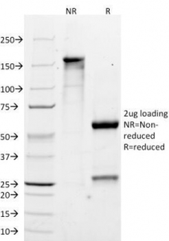
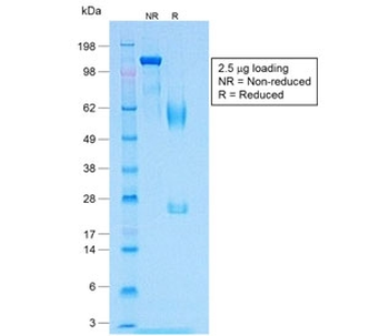
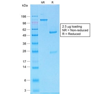
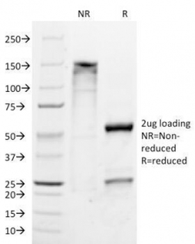
SDS-PAGE Analysis of Purified, BSA-Free MART-1 Antibody (clone M2-7C10). Confirmation of Integrity and Purity of the Antibody.
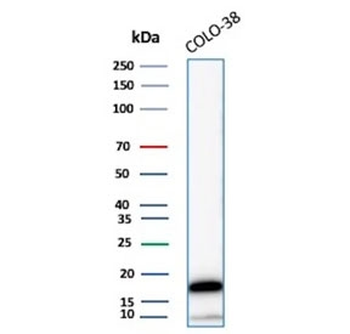
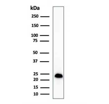
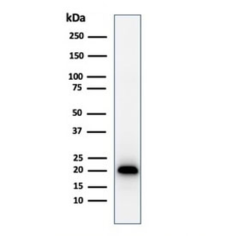
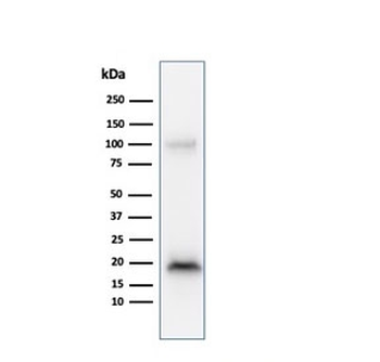
Western blot testing of human COLO-38 cell lysate with MART-1 antibody (clone M2-7C10). Expected molecular weight ~20 kDa.
- Item 1 of 5
- Item 1 of 4
- Item 1 of 4
- Item 1 of 4
MART-1 / Melan-A Antibody [orb248349]
FACS, IF, IHC-P, WB
Human, Mouse, Rat
Mouse
Monoclonal
Unconjugated
20 μg - Item 1 of 4