You have no items in your shopping cart.
Cart summary
Item 1 of 7
Item 1 of 7
KLRC1 Antibody
Catalog Number: orb1271011

| Catalog Number | orb1271011 |
|---|---|
| Category | Antibodies |
| Description | KLRC1 Antibody |
| Target | KLRC1 |
| Clonality | Polyclonal |
| Isotype | Rabbit Ig |
| Conjugation | Unconjugated |
| Reactivity | Human |
| Form/Appearance | Liquid |
| Concentration | batch dependent |
| Buffer/Preservatives | Supplied in PBS with 0.09% (W/V) sodium azide. |
| Purification | This antibody is purified through a protein A column, followed by peptide affinity purification. |
| Immunogen | This KLRC1 antibody is generated from rabbits immunized with a KLH conjugated synthetic peptide between 180-206 amino acids from the C-terminal region of human KLRC1. |
| UniProt ID | P26715 |
| MW | 26 kDa |
| Tested applications | FC, IF, IHC-P, WB |
| Application notes | For IF starting dilution is: 1:25For FACS starting dilution is: 1:25For WB starting dilution is: 1:1000For IHC-P starting dilution is: 1:50~100 |
| Antibody Type | Primary Antibody |
| Storage | Maintain refrigerated at 2-8°C for up to 2 weeks. For long term storage store at -20°C in small aliquots to prevent freeze-thaw cycles. |
| Alternative names | NKG2-A/NKG2-B type II integral membrane protein, C Read more... |
| Note | For research use only |
| NCBI | P26715 |
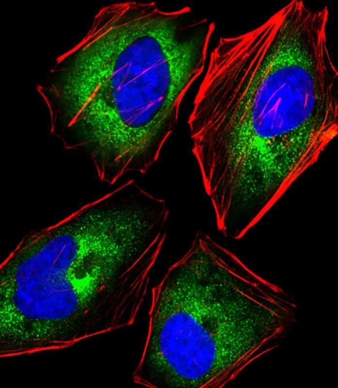
Immunofluorescent analysis of 4% paraformaldehyde-fixed, 0.1% Triton X-100 permeabilized HeLa (human cervical epithelial adenocarcinoma cell line) cells labeling Pdx1 with antibody at 1/25 dilution, followed by 488-conjugated goat anti-rabbit IgG secondary antibody at 1/200 dilution (green). Immunofluorescence image showing cytoplasm staining on HeLa cell line. Cytoplasmic actin is detected with 554 Phalloidin at 1/100 dilution (red). The nuclear counter stain is DAPI (blue).
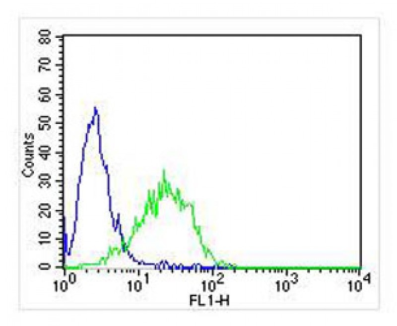
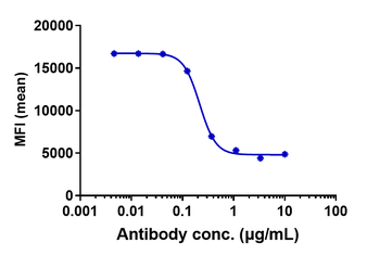
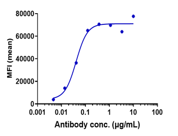
Overlay histogram showing Jurkat cells stained with Antibody (green line). The cells were fixed with 2% paraformaldehyde (10 min). The cells were then icubated in 2% bovine serum albumin to block non-specific protein-protein interactions followed by the antibody (1:25 dilution) for 60 min at 37°C. The secondary antibody used was Goat-Anti-Rabbit IgG, Conjugated Highly Cross-Adsorbed (NA168821) at 1/400 dilution for 40 min at 37°C. Isotype control antibody (blue line) was rabbit IgG (1ug/1x10^6 cells) used under the same conditions. Acquisition of > 10000 events was performed.

Overlay histogram showing Jurkat cells stained with Antibody (green line). The cells were fixed with 2% paraformaldehyde (10 min). The cells were then icubated in 2% bovine serum albumin to block non-specific protein-protein interactions followed by the antibody (1:25 dilution) for 60 min at 37°C. The secondary antibody used was Goat-Anti-Rabbit IgG, Conjugated Highly Cross-Adsorbed (NA168821) at 1/400 dilution for 40 min at 37°C. Isotype control antibody (blue line) was rabbit IgG (1ug/1x10^6 cells) used under the same conditions. Acquisition of > 10000 events was performed.
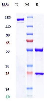
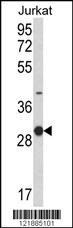
Western Blot at 1:2000 dilution + Jurkat whole cell lysate Lysates/proteins at 20 ug per lane.

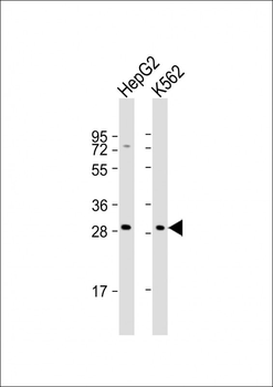
Western Blot at 1:2000 dilution Lane 1: HepG2 whole cell lysates Lane 2: K562 whole cell lysates Lysates/proteins at 20 ug per lane.
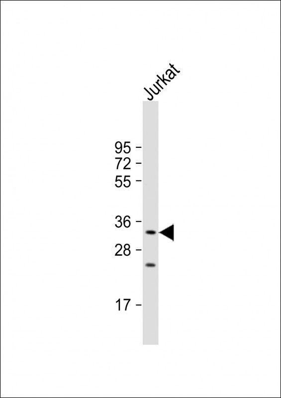
Western blot analysis of KLRC1 Antibody in Jurkat cell line lysates (35 ug/lane)
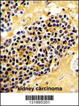
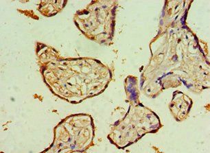
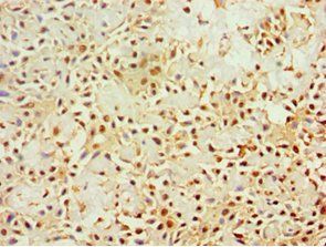
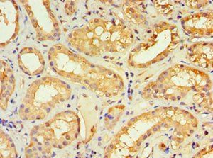
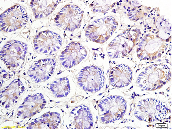
Formalin-fixed and paraffin-embedded human kidney carcinoma with KLRC1 Antibody, which was peroxidase-conjugated to the secondary antibody, followed by DAB staining.
- Item 1 of 6
KLRC1 Antibody (C-term) [orb1928387]
FC, IF, IHC-P, WB
Human
Rabbit
Polyclonal
Unconjugated
100 μl, 50 μl - Item 1 of 5
Anti-NKG2A / CD94 Reference Antibody (monalizumab) [orb1818071]
ELISA, FC
Human, Monkey, Primate
Monoclonal
Unconjugated
100 μg - Item 1 of 3
- Item 1 of 1
NKG2A Rabbit Polyclonal Antibody [orb13611]
ELISA, IF, IHC-Fr, IHC-P
Mouse, Rat
Mouse
Rabbit
Polyclonal
Unconjugated
100 μl, 50 μl, 200 μl - Item 1 of 2