You have no items in your shopping cart.
Cart summary
Item 1 of 11
Item 1 of 11
IRF3 Antibody
Catalog Number: orb2636911

| Catalog Number | orb2636911 |
|---|---|
| Category | Antibodies |
| Description | In the absence of viral infection, IRF3 is maintained as a monomer in an autoinhibited state. Phosphorylation by TBK1 and IKBKE disrupts this autoinhibition leading to the liberation of the DNA-binding and dimerization activities and its nuclear localization where it can activate type I IFN and ISG genes. [UniProt] |
| Clonality | Monoclonal |
| Species/Host | Mouse |
| Isotype | Mouse IgG2b, kappa |
| Conjugation | Unconjugated |
| Reactivity | Human |
| Buffer/Preservatives | 0.2 mg/ml in 1X PBS with 0.1 mg/ml rAlbumin (US sourced), 0.05% sodium azide |
| Purity | Protein A/G affinity |
| Immunogen | Recombinant full-length human IRF3 protein was used as the immunogen for the IRF3 antibody. |
| UniProt ID | Q14653 |
| Tested applications | FACS, IF, WB |
| Dilution range | Flow cytometry: 1-2ug/million cells,Immunofluorescence: 1-2ug/ml,Western blot: 1-2ug/ml |
| Application notes | Optimal dilution of the IRF3 antibody should be determined by the researcher. |
| Antibody Type | Primary Antibody |
| Clone Number | PCRP-IRF3-6C8 |
| Formula | 0.2 mg/ml in 1X PBS with 0.1 mg/ml BSA (US sourced), 0.05% sodium azide |
| Storage | Maintain refrigerated at 2-8°C for up to 2 weeks. For long term storage store at -20°C in small aliquots to prevent freeze-thaw cycles. |
| Hazard Information | This IRF3 antibody is available for research use only. |
| Note | For research use only |

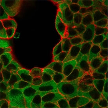
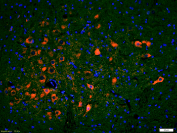
Immunofluorescent staining of human MCF-7 cells using IRF3 antibody (green, clone PCRP-IRF3-6C8) and phalloidin (red).
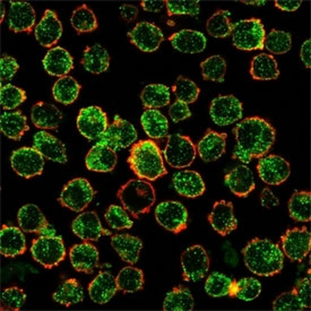
Immunofluorescent staining of human K562 cells using IRF3 antibody (green, clone PCRP-IRF3-6C8) and phalloidin (red).
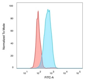
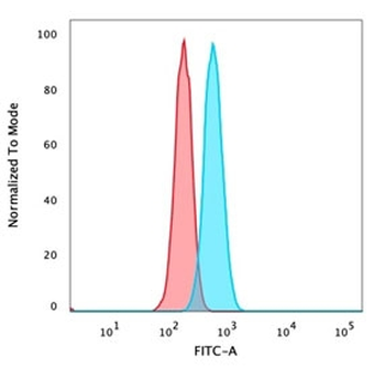
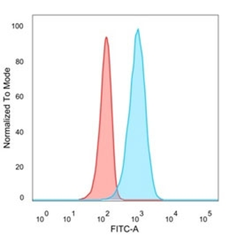
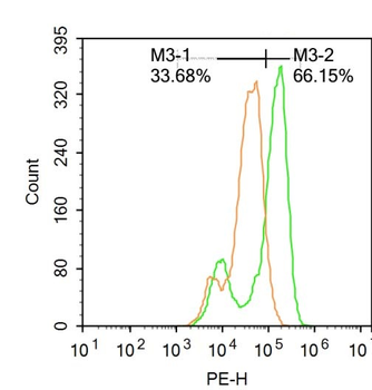
FACS staining of PFA-fixed human MCF-7 cells with IRF3 antibody (blue, clone PCRP-IRF3-6C8), and unstained cells (red).
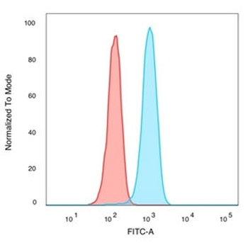
FACS staining of PFA-fixed human K562 cells with IRF3 antibody (blue, clone PCRP-IRF3-6C8), and unstained cells (red).

FACS staining of PFA-fixed human HeLa cells with IRF3 antibody (blue, clone PCRP-IRF3-6C8), and unstained cells (red).

FACS staining of PFA-fixed human U87 cells with IRF3 antibody (blue, clone PCRP-IRF3-6C8), and unstained cells (red).
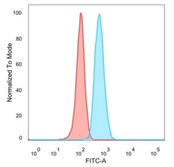
FACS staining of PFA-fixed human Raji cells with IRF3 antibody (blue, clone PCRP-IRF3-6C8), and unstained cells (red).
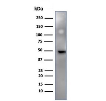
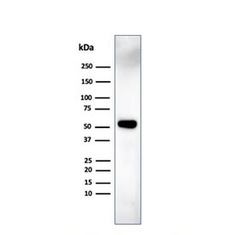
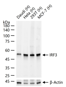
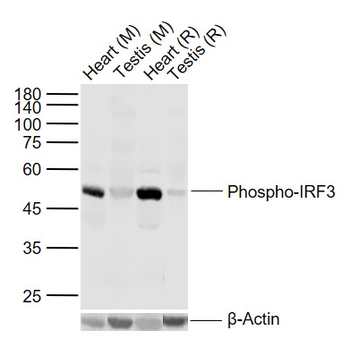
Western blot testing of human HeLa cell lysate using IRF3 antibody (clone PCRP-IRF3-6C8). Predicted molecular weight ~47 kDa.

Western blot testing of human MCF7 cell lysate using IRF3 antibody (clone PCRP-IRF3-6C8). Predicted molecular weight ~47 kDa.
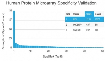
Analysis of HuProt (TM) microarray containing more than 19000 full-length human proteins using IRF3 antibody (clone PCRP-IRF3-6C8). These results demonstrate the foremost specificity of the PCRP-IRF3-6C8 mAb. Z- and S- score: The Z-score represents the strength of a signal that an antibody (in combination with a fluorescently-tagged anti-IgG secondary Ab) produces when binding to a particular protein on the HuProt (TM) array. Z-scores are described in units of standard deviations (SD's) above the mean value of all signals generated on that array. If the targets on the HuProt (TM) are arranged in descending order of the Z-score, the S-score is the difference (also in units of SD's) between the Z-scores. The S-score therefore represents the relative target specificity of an Ab to its intended target.
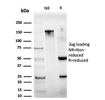
SDS-PAGE analysis of purified, BSA-free IRF3 antibody (PCRP-IRF3-6C8) as confirmation of integrity and purity.
- Item 1 of 11
- Item 1 of 10
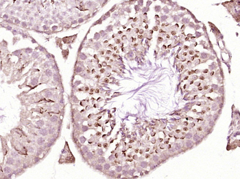
IRF3 Recombinant Rabbit Monoclonal Antibody [orb500087]
IF, IHC-Fr, IHC-P, WB
Rat
Human, Mouse
Rabbit
Recombinant
Unconjugated
25 μl, 50 μl, 100 μl - Item 1 of 6
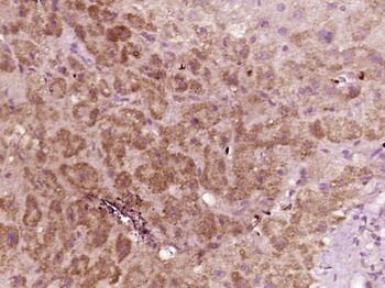
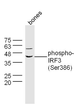
Phospho-IRF3 (Ser386) Rabbit Polyclonal Antibody [orb1860]
FC, IF, IHC-Fr, IHC-P, WB
Bovine, Canine, Porcine, Rabbit, Rat
Human, Mouse
Rabbit
Polyclonal
Unconjugated
50 μl, 100 μl, 200 μl - Item 1 of 5
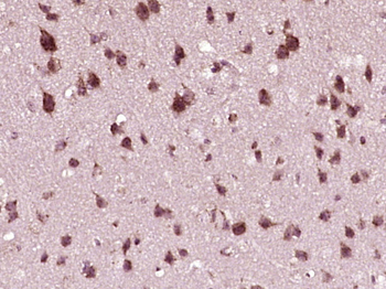
Phospho-IRF3 (Ser396) Rabbit Polyclonal Antibody [orb6225]
IF, IHC-Fr, IHC-P, WB
Bovine, Canine, Porcine, Sheep
Human, Mouse, Rat
Rabbit
Polyclonal
Unconjugated
50 μl, 100 μl, 200 μl - Item 1 of 6