You have no items in your shopping cart.
Description
Images & Validation
−Item 1 of 1
| Tested Applications | FC, FLISA, IF |
|---|---|
| Dilution Range | FLISA: 1:10,000 - 1:50,000, FC: 1:500 - 1:2,500, IF: 1:1,000 - 1:5,000 |
| Reactivity | Human |
| Application Notes |
Key Properties
−| Antibody Type | Secondary Antibody |
|---|---|
| Host | Rabbit |
| Clonality | Polyclonal |
| Isotype | IgG |
| Immunogen | Human IgG whole molecule |
| Purity | This product is an IgG fraction antibody purified from monospecific antiserum by a multi-step process which includes delipidation, salt fractionation and ion exchange chromatography followed by extensive dialysis against the buffer stated above. Assay by immunoelectrophoresis resulted in a single precipitin arc against anti-fluorescein, anti-Rabbit Serum, Human IgG and Human Serum. |
| Conjugation | FITC |
Storage & Handling
−| Storage | Store vial at 4° C prior to restoration. For extended storage aliquot contents and freeze at -20° C or below. Avoid cycles of freezing and thawing. Centrifuge product if not completely clear after standing at room temperature. This product is stable for several weeks at 4° C as an undiluted liquid. Dilute only prior to immediate use. |
|---|---|
| Form/Appearance | Lyophilized |
| Buffer/Preservatives | Buffer: 0.02 M Potassium Phosphate, 0.15 M Sodium Chloride, pH 7.2 |
| Concentration | 10 mg/mL |
| Expiration Date | 12 months from date of receipt. |
| Disclaimer | For research use only |
Alternative Names
−rabbit anti-Human IgG Fluorescein Conjugated Antibody, rabbit anti-Human IgG FITC Conjugated Antibody, rabbit anti-Human IgG Antibody Fluorescein Conjugation
Similar Products
−- Item 1 of 2
Human IgG (H&L) Antibody Fluorescein Conjugated [orb347213]
DOT, FC, FLISA, IF, WB
Human
Goat
Polyclonal
FITC
2 mg - Item 1 of 1
F(ab')2 Human IgG (H&L) Antibody Fluorescein Conjugated Pre-Adsorbed [orb348182]
DOT, FC, FLISA, IF
Human
Goat
Polyclonal
FITC
1 mg - Item 1 of 1
Fab Human IgG (H&L) Antibody Fluorescein Conjugated [orb348459]
DOT, FC, FLISA, IF, WB
Human
Goat
Polyclonal
FITC
1 mg - Item 1 of 1
Human IgG (H&L) Antibody Fluorescein Conjugated [orb346651]
FC, FLISA, IF
Human
Mouse
Polyclonal
FITC
20 mg - Item 1 of 1
Human IgG IgA IgM (H&L) Antibody Fluorescein Conjugated [orb347190]
DOT, FC, FLISA, IF, WB
Human
Goat
Polyclonal
FITC
2 mg

Quality Guarantee
Explore bioreagents carefree to elevate your research. All our products are rigorously tested for performance. If a product does not perform as described on its datasheet, our scientific support team will provide expert troubleshooting, a prompt replacement, or a refund. For full details, please see our Terms & Conditions and Buying Guide. Contact us at [email protected].
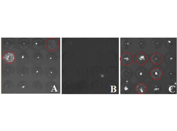
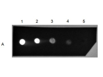
IgG Secretion. A) ARH-77, B) CCL-119, C) SA13. Both ARH-77 and SA13 cell lines display high fluorescence readouts while CCL-119 shows little fluorescence. Red circles correspond to MBs that contained cells and displayed high levels of immunoprecipitation. Light fluorescent spot in (B) is attributed to a reflection off an out of focus bubble on MB surface. By day 1 the fluorescent images showed that some wells took on a speckled appearance that continued to intensify over time (Fig. 1). By day 4 distinct differences could be discerned between the three cell lines. The fluorescence speckle pattern signifies detection of immunoprecipitation (IP) between secreted IgG and FITC labeled α-IgG added to the cell culture media.
Documents Download
Datasheet
Product Information
Request a Document
Protocol Information
FC
Flow Cytometry
IF
Immunofluorescence
Human IgG (H&L) Antibody Fluorescein Conjugated (orb346656)

- 0.0
Based on 0 reviews
Participating in our Biorbyt product reviews program enables you to support fellow scientists by sharing your firsthand experience with our products.
Login to Submit a ReviewAvailable Sizes
Select a size below
Free Secondary Antibody (20 ul)0/0
Please add an antibody product to your cart first.