You have no items in your shopping cart.
Cart summary
Item 1 of 4
Item 1 of 4
HMGA1 Antibody
Catalog Number: orb1262374
| Catalog Number | orb1262374 |
|---|---|
| Category | Antibodies |
| Description | HMGA1 Antibody |
| Species/Host | Rabbit |
| Clonality | Polyclonal |
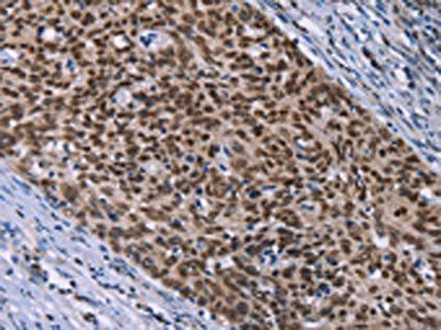
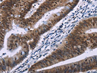
| Tested applications | FC, IF, WB |
| Predicted Reactivity | Hamster, Mouse, Rat |
| Reactivity | Human |
| Isotype | Rabbit Ig |
| Immunogen | This HMGA1 antibody is generated from rabbits immunized with a KLH conjugated synthetic peptide between 64-93 amino acids from the C-terminal region of human HMGA1. |
| Concentration | batch dependent |
| Dilution range | For WB starting dilution is: 1:1000For IF starting dilution is: 1:10~50For FACS starting dilution is: 1:10~50 |
| Form/Appearance | Liquid |
| Conjugation | Unconjugated |
| MW | 12 kDa |
| Target | HMGA1 |
| UniProt ID | P17096 |
| NCBI | P17096 |
| Storage | Store at 4°C for three months and -20°C, stable for up to one year. As with all antibodies care should be taken to avoid repeated freeze thaw cycles. Antibodies should not be exposed to prolonged high temperatures. |
| Buffer/Preservatives | Supplied in PBS with 0.09% (W/V) sodium azide. |
| Alternative names | High mobility group protein HMG-I/HMG-Y, HMG-I(Y), Read more... |
| Note | For research use only |
| Application notes | For WB starting dilution is: 1:1000For IF starting dilution is: 1:10~50For FACS starting dilution is: 1:10~50 |
| Expiration Date | 12 months from date of receipt. |
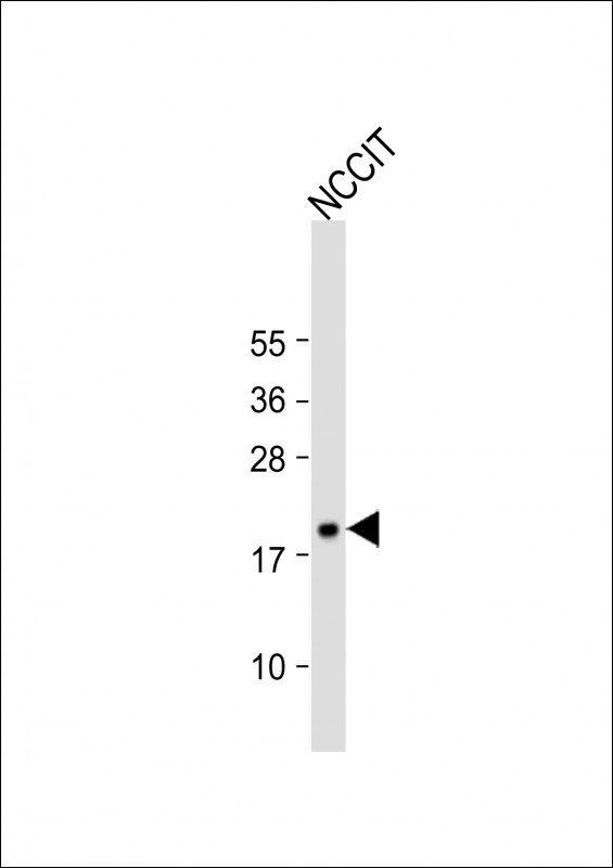
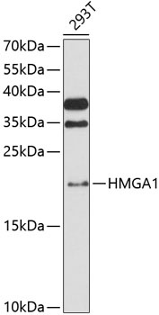
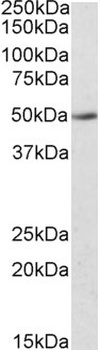
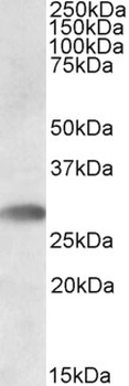
Western Blot at 1:1000 dilution + NCCIT whole cell lysate Lysates/proteins at 20 ug per lane.
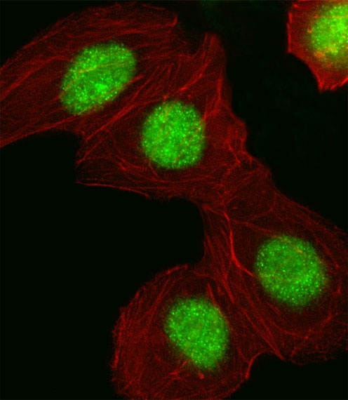
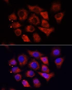
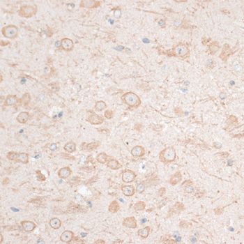
Fluorescent image of A549 cell stained with HMGA1 Antibody.A549 cells were fixed with 4% PFA (20 min), permeabilized with Triton X-100 (0.1%, 10 min), then incubated with HMGA1 primary antibody (1:25). For secondary antibody, Alexa Fluor 488 conjugated donkey anti-rabbit antibody (green) was used (1:400).Cytoplasmic actin was counterstained with Alexa Fluor 555 (red) conjugated Phalloidin (7 units/ml).HMGA1 immunoreactivity is localized to Nucleus significantly.
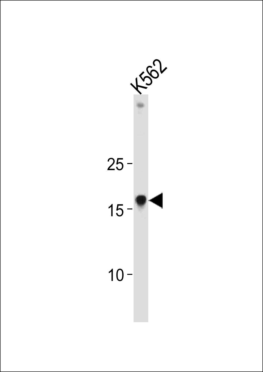
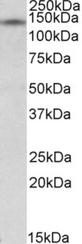
Western blot analysis in K562 cell line lysates (35 ug/lane).
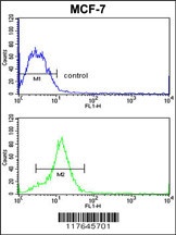
Flow cytometric analysis of MCF-7 cells (bottom histogram) compared to a negative control cell (top histogram). FITC-conjugated goat-anti-rabbit secondary antibodies were used for the analysis.
- Item 1 of 3
- Item 1 of 3
- Item 1 of 2
- Item 1 of 3
HMGA1 Antibody [orb1249518]
ELISA, IHC, WB
Bovine, Canine, Mouse, Porcine, Rat
Human
Goat
Polyclonal
Unconjugated
0.1 mg - Item 1 of 3


Submit a review
Filter by Rating

- 5 stars

- 4 stars
- 3 stars
- 2 stars
- 1 stars