You have no items in your shopping cart.
Cart summary
Item 1 of 3
Item 1 of 3
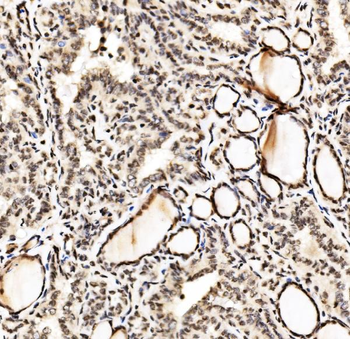
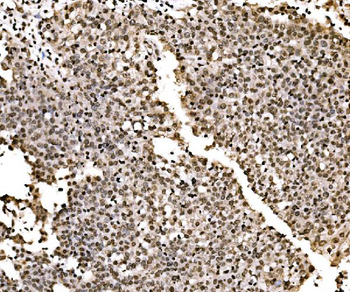
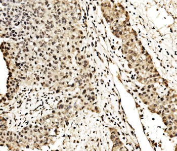
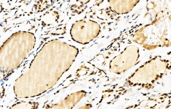
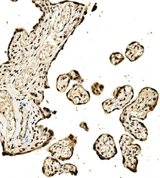
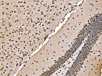
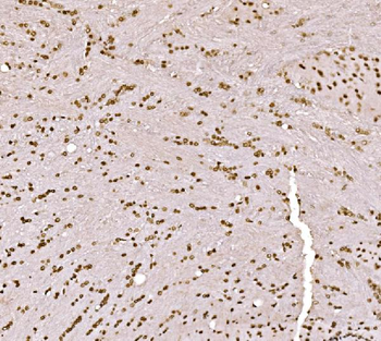
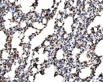
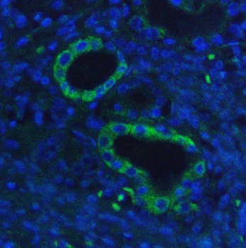
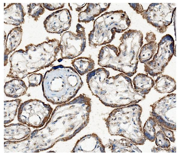
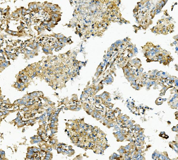
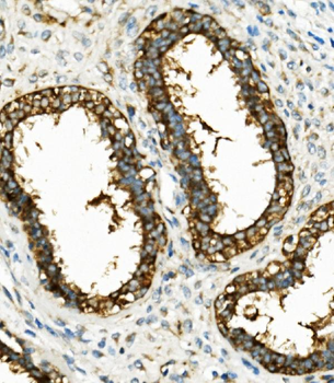
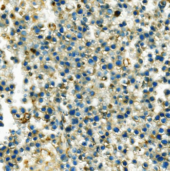
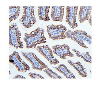
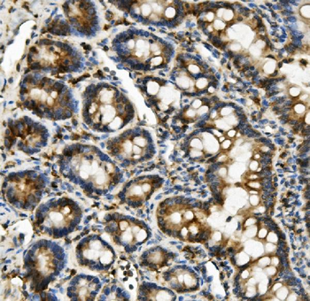
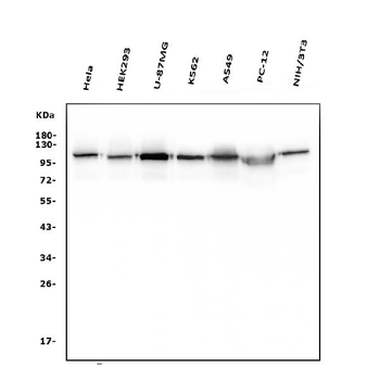
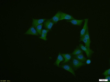
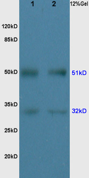
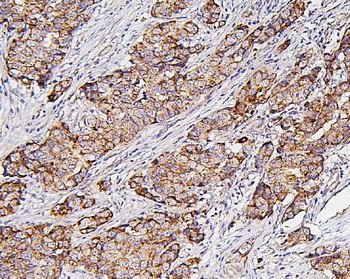
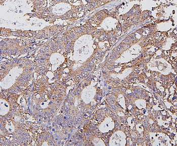
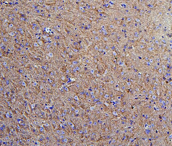
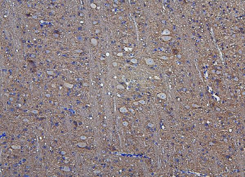
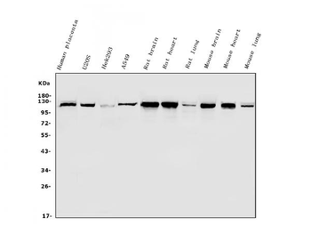
Hexokinase Antibody
Catalog Number: orb750579

| Catalog Number | orb750579 |
|---|---|
| Category | Antibodies |
| Description | HXK2 antibody |
| Clonality | Polyclonal |
| Species/Host | Rabbit |
| Isotype | Antiserum |
| Conjugation | Unconjugated |
| Form/Appearance | Lyophilized |
| Concentration | 80 mg/mL |
| Buffer/Preservatives | 0.01% (w/v) Sodium Azide |
| Purity | This product was prepared from monospecific antiserum by a delipidation and defibrination. Assay by immunoelectrophoresis resulted in a single precipitin arc against anti-rabbit serum, purified and partially purified Hexokinase [Yeast]. Cross reactivity against Hexokinase from other tissues and species may occur but have not been specifically determined. |
| Immunogen | Hexokinase [Yeast] |
| UniProt ID | P04806 |
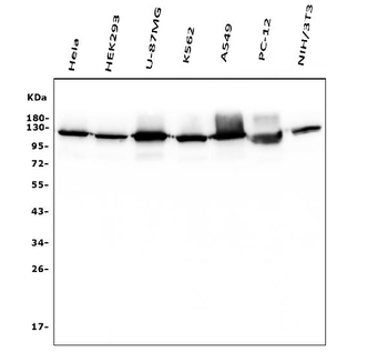
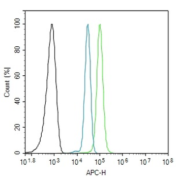
| Tested applications | ELISA, WB |
| Dilution range | ELISA: 1:5,000 - 1:25,000, WB: 1:500 - 1:3,000 |
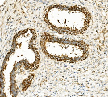
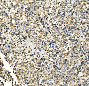
| Application notes | Anti-Hexokinase is suitable for use in ELISA, western blot, and immunohistochemistry. Specific conditions for reactivity should be optimized by the end user. |
| Antibody Type | Primary Antibody |
| Storage | Store vial at 4° C prior to restoration. For extended storage aliquot contents and freeze at -20° C or below. Avoid cycles of freezing and thawing. Centrifuge product if not completely clear after standing at room temperature. This product is stable for several weeks at 4° C as an undiluted liquid. Dilute only prior to immediate use. |
| Alternative names | rabbit anti-Hexokinase Antibody, DKFZp686M1669 ant Read more... |
| Note | For research use only |
| NCBI | CAA96973.1 |
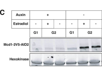
Effects of cohesin depletion and tT(AGU)C deletion on silencing establishment.(A) SIR3-EBD strains (JRY12269, JRY12270) and SIR3-EBD strains with seamless deletion of tT(AGU)C (JRY12267; JRY12268) were arrested in G1 with α factor, then split, with half the culture receiving estradiol and half receiving ethanol. Samples were collected after 3 hr for RT-qPCR, with each sample normalized to its own pre-estradiol value. (B) Cells with MCD1-AID (JRY12560, JRY12561) were arrested in G1 with α factor, then split, with half receiving auxin and the other half receiving DMSO (solvent control). After 30 min, each culture was further split, with half receiving estradiol and the other half ethanol. All cultures were released to G2/M by addition of protease and nocodazole. Cells were collected after 3 hr for RT-qPCR, with each sample normalized to its own pre-estradiol value. (C) Immunoblot analysis showing Mcd1-AID depletion for experiment described in (B).
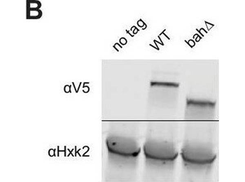
Nucleosome binding was required for spread, but not recruitment, of Sir3 to regions of heterochromatin. (A) Schematic of Sir3 protein domains. (B) Protein immunoblotting in strains expressing Sir3 (no tag, JRY11699), Sir3-3xV5 (JRY12601), and sir3-bah, àÜ, Äì3xV5 (JRY13621). Top row are 3xV5-tagged Sir3 proteins, and bottom row is the loading control Hxk2. The unedited blot is in Figure 2, Äîsource data 1. (C) RT-qPCR of HMLŒ±2 and HMRa1 mRNA, normalized to ACT1 mRNA, in strains expressing SIR3-M.ECOGII (JRY12840, JRY13027), sir3, àÜ::M.ECOGII (JRY13029, JRY13030), and sir3-bah, àÜ-M.ECOGII (JRY13438, JRY13439). Data are the average of three biological replicates, and bars mark one standard deviation. (D) Aggregate methylation results at HML (top) and HMR (bottom) from long-read Nanopore sequencing of sir3, àÜ::M.ECOGII (JRY13029, JRY13030), SIR3-M.ECOGII (JRY12840, JRY13027), and sir3-bah, àÜ-M.ECOGII (JRY13438). Plots are as described in Figure 1D. (E) Single-read plots from long-read Nanopore sequencing of sir3-bah, àÜ-M.ECOGII (JRY13438) at HML (top) and HMR (bottom). Plots are as described in Figure 1E. (F) Aggregate methylation results at four representative telomeres (1 L, 2 L, 4 R, and 11 R) from long-read Nanopore sequencing of the same strains as D. Shown are 15 kb windows of each telomere. Plots are as described in Figure 1D. (G) Single-read plots from long-read Nanopore sequencing of SIR3-M.ECOGII (JRY13027) and sir3-bah, àÜ-M.ECOGII (JRY13438) at two representative telomeres (1 L and 2 L). Shown are 10 kb windows of each telomere. Figure 2, Äîsource data 1.Uncropped protein immunoblot of Sir3 mutants. Uncropped protein immunoblot of Sir3 mutants. DIP-seq of SIR3-M.ECOGII (top row, JRY13027), sir3, àÜ::M.ECOGII (middle row, JRY13030), and sir3-bah, àÜ-M.ECOGII (bottom row, JRY13438).Input results are plotted but not visible due to the strong DIP-seq signals. (A) Shown are 10 kb regions centered at HML (left) and HMR (right). (B) Shown are 10 kb regions at two representative telomeres (1 L and 2 L). Methylation by Sir3, ÄìM.EcoGII and sir3-bah, àÜ-M.EcoGII at all 32 telomeres. Aggregate methylation results from long-read Nanopore sequencing of the same strains as Figure 2D. Shown are 15 kb windows of each telomere. Plots are as described in Figure 1D. Highlighted in yellow are windows shorter than 15 kb due to discrepancies between the S288C and W303 genomes.
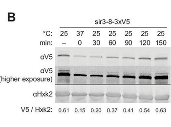
Repression of HML and HMR preceded heterochromatin maturation.(A) Schematic of temperature-shift time course with sir3-8-M.ECOGII. (B) Protein immunoblotting in a strain expressing sir3-8-3xV5 (JRY13467) constitutively at 25¬∞C (first lane), constitutively at 37¬∞C (second lane), and at 30, 60, 90, 120, and 150 min after a shift to 25¬∞C. Top row is 3xV5-tagged sir3-8 protein, the middle row is the same as the top row but at a higher exposure, and the bottom row is the loading control Hxk2. The unedited blot is in. (C) Aggregate methylation results at HML (top) and HMR (bottom) from long-read Nanopore sequencing of strains expressing sir3-8-M.ECOGII (JRY13114) grown constitutively at 25 or 37¬∞C. Plots are as described in Figure 1D. (D) Aggregate methylation results at HML (top) and HMR (bottom) from long-read Nanopore sequencing of a strain expressing sir3-8-M.ECOGII (JRY13134) grown constitutively at 25¬∞C (dotted gray line) and collected at 0, 15, 45, and 90 min after a temperature switch from 37 to 25¬∞C. (E) RT-qPCR of HMLŒ±2 (left) and HMRa1 (right) mRNA in strains expressing SIR3-M.ECOGII (black, JRY13027, JRY12840), sir3, àÜ::M.ECOGII (green, JRY13029, JRY13030), or sir3-8-M.ECOGII (purple, JRY13114, JRY13134) collected at 0, 30, 60, 90, 120, and 150 min after a temperature switch from 37 to 25¬∞C. Points are the average of three biological replicates and bars mark one standard deviation. DIP-seq of sir3-8-M.ECOGII (JRY13114). Shown are 10 kb regions centered at HML (left) and HMR (right). Cells were grown constitutively at either 25 or 37¬∞C. Input results are plotted but not visible due to the strong DIP-seq signals. Nanopore sequencing over temperature switch time course (biological replicate).Aggregate methylation results at HML (top) and HMR (bottom) from long-read Nanopore sequencing of a strain expressing sir3-8-M.ECOGII (JRY13114) grown constitutively at 25¬∞C (dotted gray line) and collected at 0, 15, 45, and 90 min after a temperature switch from 37 to 25¬∞C.
- Item 1 of 15
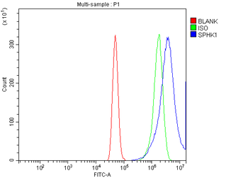
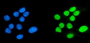
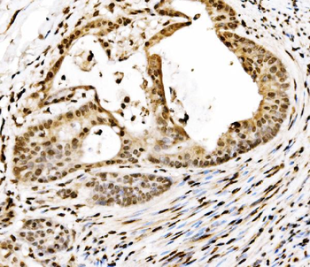
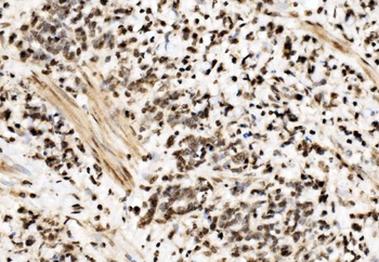
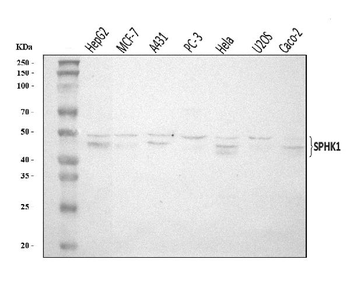
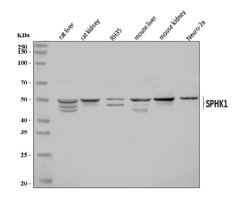
Anti-SPHK1 Antibody [orb763192]
ELISA, FC, ICC, IF, IHC, WB
Human, Mouse, Rat
Rabbit
Polyclonal
Unconjugated
100 μg, 10 μg - Item 1 of 10
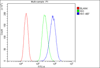
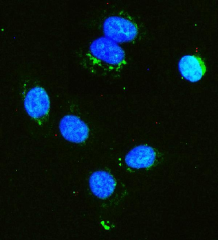
Anti-Hexokinase 1/HK1 Antibody (monoclonal, 4B7) [orb654274]
FC, ICC, IF, IHC, IHC-Fr, WB
Human, Mouse, Rat
Mouse
Monoclonal
Unconjugated
10 μg, 100 μg - Item 1 of 7
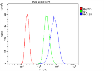
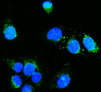
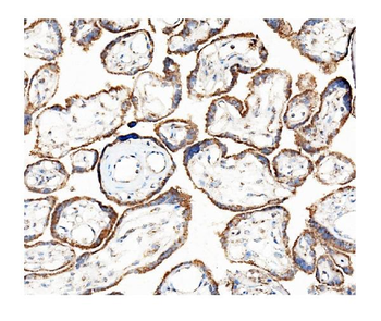
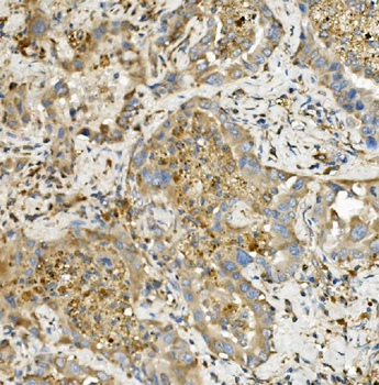
Anti-Hexokinase 1/HK1 Antibody (monoclonal, 2I4) [orb654273]
FC, ICC, IF, IHC, IHC-Fr, WB
Human, Mouse, Rat
Mouse
Monoclonal
Unconjugated
10 μg, 100 μg - Item 1 of 4
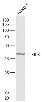
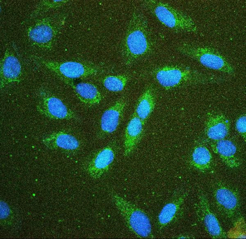
GLK Rabbit Polyclonal Antibody [orb13428]
FC, ICC, WB
Canine, Equine, Gallus, Mouse, Porcine, Rabbit
Human, Rat
Rabbit
Polyclonal
Unconjugated
50 μl, 100 μl, 200 μl - Item 1 of 6
Hexokinase 1/HK1 Antibody [orb570337]
ELISA, ICC, IF, IHC, WB
Human, Mouse, Rat
Rabbit
Polyclonal
Unconjugated
10 μg, 100 μg