You have no items in your shopping cart.
Cart summary
Item 1 of 6
Item 1 of 6
GLS Antibody
Catalog Number: orb1271010

| Catalog Number | orb1271010 |
|---|---|
| Category | Antibodies |
| Description | GLS Antibody |
| Target | GLS |
| Clonality | Polyclonal |
| Isotype | Rabbit Ig |
| Conjugation | Unconjugated |
| Reactivity | Human, Mouse, Rat |
| Form/Appearance | Liquid |
| Concentration | batch dependent |
| Buffer/Preservatives | Supplied in PBS with 0.09% (W/V) sodium azide. |
| Immunogen | This GLS antibody is generated from rabbits immunized with a KLH conjugated synthetic peptide between 516-545 amino acids from the C-terminal region of human GLS. |
| UniProt ID | O94925 |
| MW | 73 kDa |
| Tested applications | IF, IHC-P, WB |
| Application notes | For WB starting dilution is: 1:2000For IHC-P starting dilution is: 1:25For IF starting dilution is: 1:25For IF starting dilution is: 1:25 |
| Antibody Type | Primary Antibody |
| Storage | Maintain refrigerated at 2-8°C for up to 2 weeks. For long term storage store at -20°C in small aliquots to prevent freeze-thaw cycles. |
| Alternative names | Glutaminase kidney isoform, mitochondrial, GLS, K- Read more... |
| Note | For research use only |
| NCBI | O94925 |
| Expiration Date | 12 months from date of receipt. |
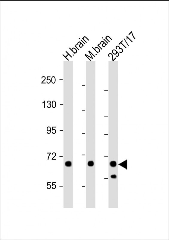
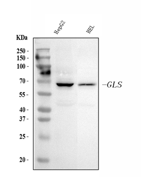
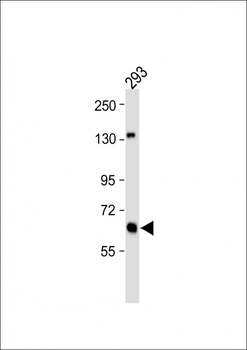
Western Blot at 1:2000 dilution Lane 1: human brain lysate Lane 2: mouse brain lysate Lane 3: 293T/17 whole cell lysate Lysates/proteins at 20 ug per lane.

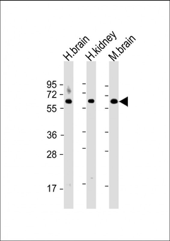
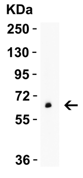
Western Blot at 1:2000 dilution Lane 1: human brain lysate Lane 2: human kidney lysate Lane 3: mouse brain lysate Lysates/proteins at 20 ug per lane.
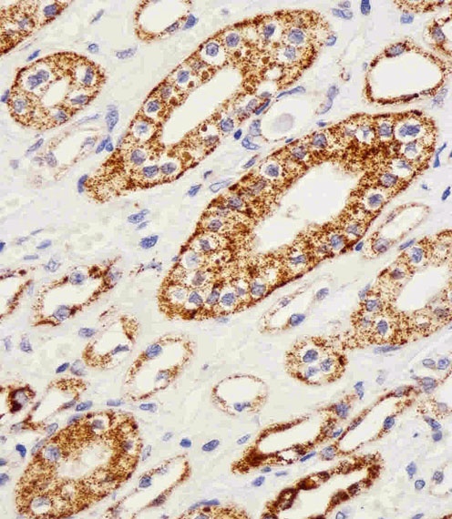
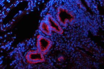
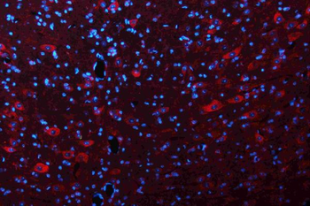
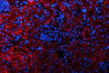
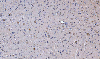
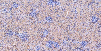
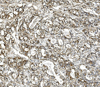
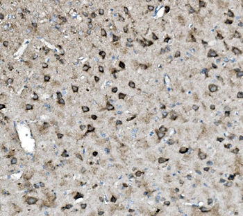
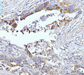
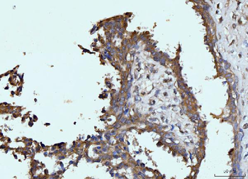
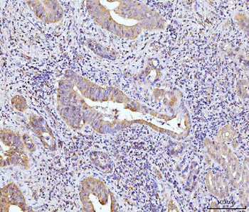
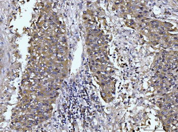
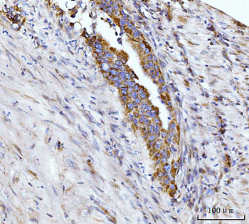
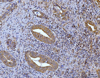
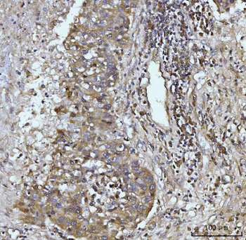
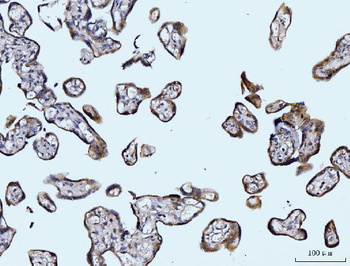
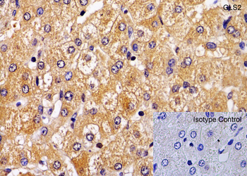
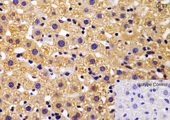
Immunohistochemical analysis of paraffin-embedded H.kidney section using GLS Antibody. Antibody was diluted at 1:25 dilution. A peroxidase-conjugated goat anti-rabbit IgG at 1:400 dilution was used as the secondary antibody, followed by DAB staining.
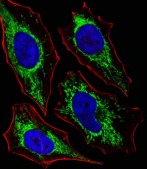
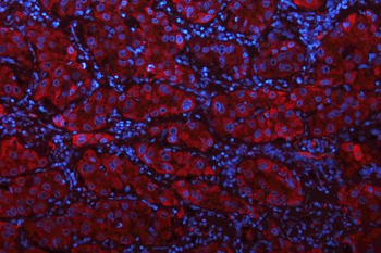
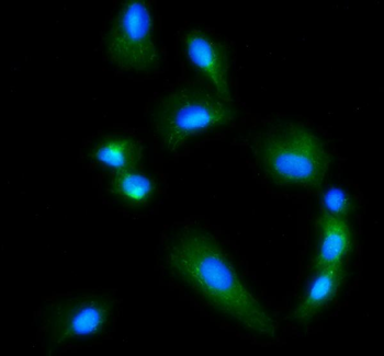
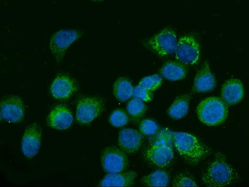
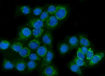
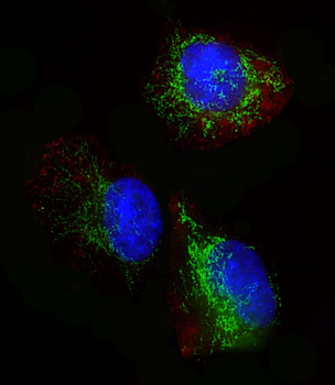
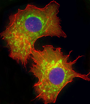
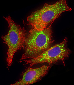
Fluorescent image of Hela cells stained with XAF1 GLS Antibody. Antibody was diluted at 1:25 dilution. An Alexa Fluor 488-conjugated goat anti-rabbit lgG at 1:400 dilution was used as the secondary antibody (green). DAPI was used to stain the cell nuclear (blue). Cytoplasmic actin was counterstained with Alexa Fluor 555 conjugated with Phalloidin (red).
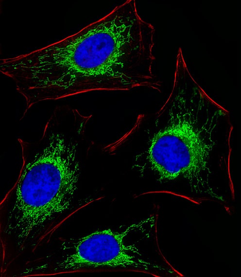
Fluorescent image of Hela cells stained with XAF1 GLS Antibody. Antibody was diluted at 1:25 dilution. An Alexa Fluor 488-conjugated goat anti-rabbit lgG at 1:400 dilution was used as the secondary antibody (green). DAPI was used to stain the cell nuclear (blue). Cytoplasmic actin was counterstained with Alexa Fluor 555 conjugated with Phalloidin (red).

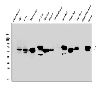
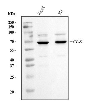
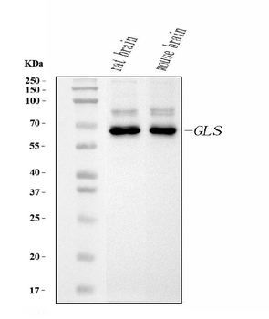
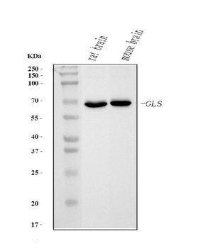
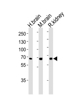
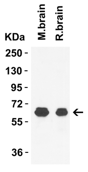
Western blot analysis of lysates from human brain, mouse brain ad rat kidney tissue lysate (from left to right), using GLS Antibody at 1:1000 at each lane.
- Item 1 of 13
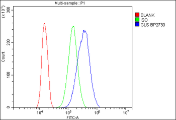
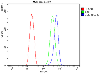
Anti-Glutaminase/GLS Antibody [orb654427]
ELISA, FC, ICC, IF, IHC, WB
Human, Monkey, Mouse, Rat
Rabbit
Polyclonal
Unconjugated
10 μg, 100 μg - Item 1 of 8
Anti-Glutaminase/GLS Antibody (monoclonal, 3G13) [orb865661]
FC, ICC, IF, IHC, WB
Human, Mouse, Rat
Mouse
Monoclonal
Unconjugated
10 μg, 100 μg - Item 1 of 7
Anti-Glutaminase/GLS Antibody (monoclonal, 9G6) [orb865660]
ICC, IF, IHC, WB
Human, Mouse, Rat
Mouse
Monoclonal
Unconjugated
10 μg, 100 μg - Item 1 of 7
GLS Antibody (C-term) [orb1928270]
FC, IF, IHC-P, WB
Human, Mouse, Rat
Rabbit
Polyclonal
Unconjugated
100 μl, 50 μl - Item 1 of 6
GLS2 Antibody [orb1239416]
ELISA, IF, IHC-P, WB
Human, Mouse, Rat
Polyclonal
Unconjugated
0.1 mg, 0.02 mg