You have no items in your shopping cart.
Cart summary
Item 1 of 4
Item 1 of 4
GLA Antibody
Catalog Number: orb1263691

| Catalog Number | orb1263691 |
|---|---|
| Category | Antibodies |
| Description | GLA Antibody |
| Species/Host | Rabbit |
| Clonality | Polyclonal |
| Tested applications | FC, IF, IHC-P, WB |
| Reactivity | Human |
| Isotype | Rabbit Ig |
| Immunogen | This GLA antibody is generated from rabbits immunized with a KLH conjugated synthetic peptide between 83-112 amino acids from the N-terminal region of human GLA. |
| Antibody Type | Primary Antibody |
| Concentration | batch dependent |
| Form/Appearance | Liquid |
| Conjugation | Unconjugated |
| MW | 49 kDa |
| Target | GLA |
| UniProt ID | P06280 |
| NCBI | P06280 |
| Storage | Maintain refrigerated at 2-8°C for up to 2 weeks. For long term storage store at -20°C in small aliquots to prevent freeze-thaw cycles. |
| Buffer/Preservatives | Supplied in PBS with 0.09% (W/V) sodium azide. |
| Alternative names | Alpha-galactosidase A, Alpha-D-galactosidase A, Al Read more... |
| Note | For research use only |
| Application notes | For WB starting dilution is: 1:1000For IHC-P starting dilution is: 1:10~50For IF starting dilution is: 1:10~50For FACS starting dilution is: 1:10~50 |
| Expiration Date | 12 months from date of receipt. |
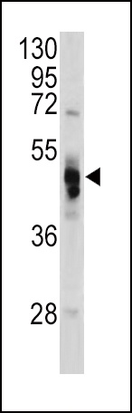
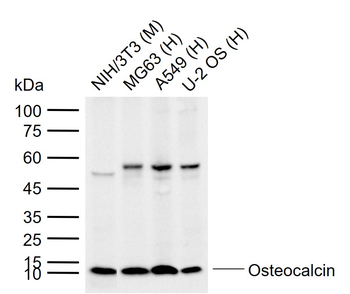
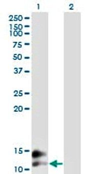
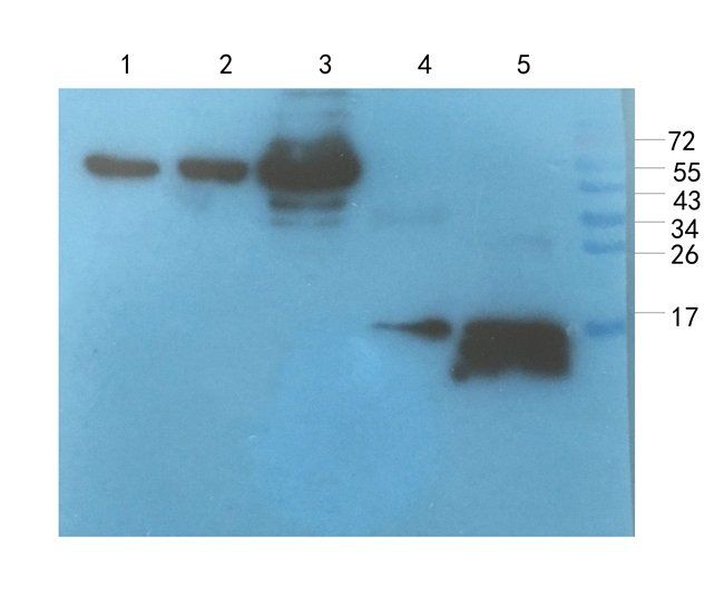
Western blot analysis of GLA antibody in Hela cell line lysates (35 ug/lane)
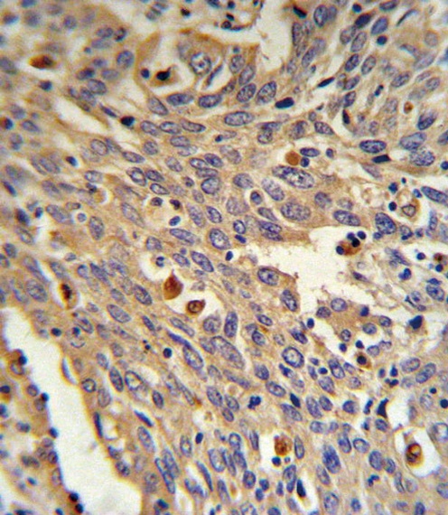
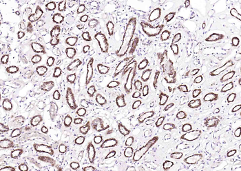
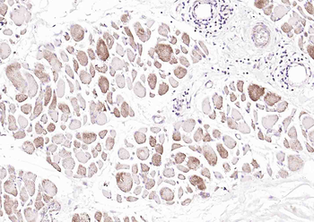
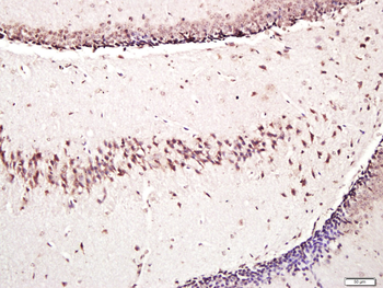
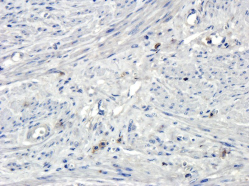
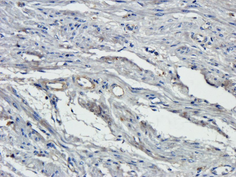
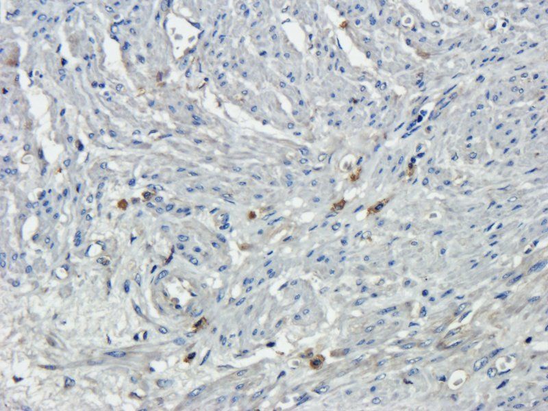
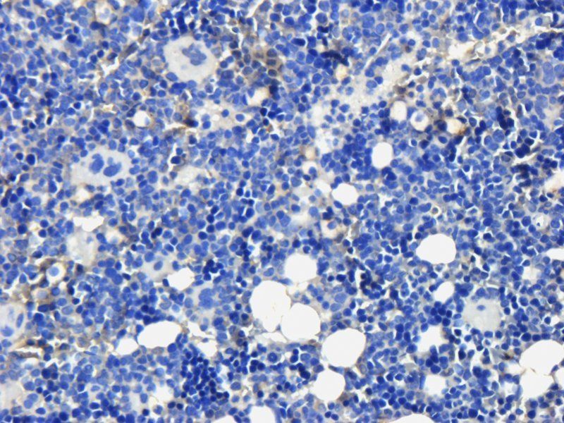
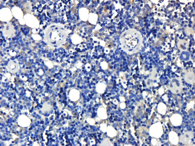
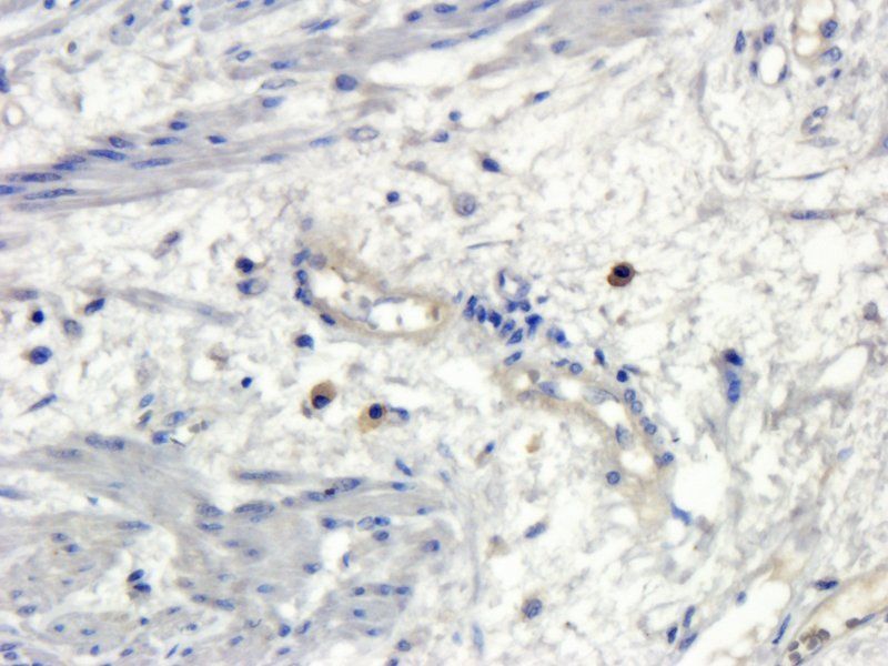
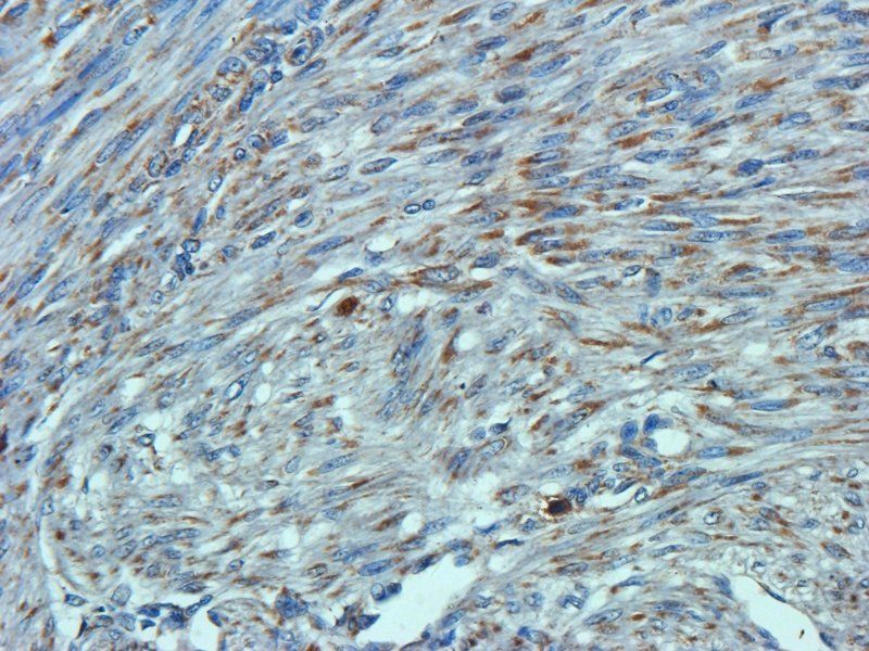
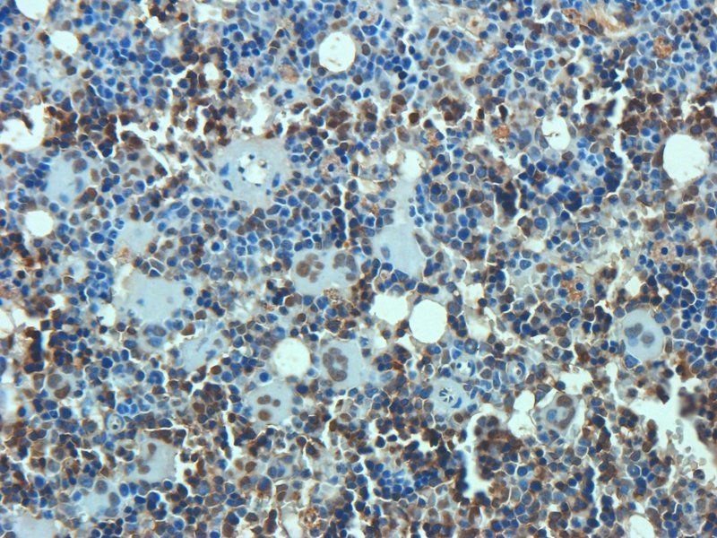
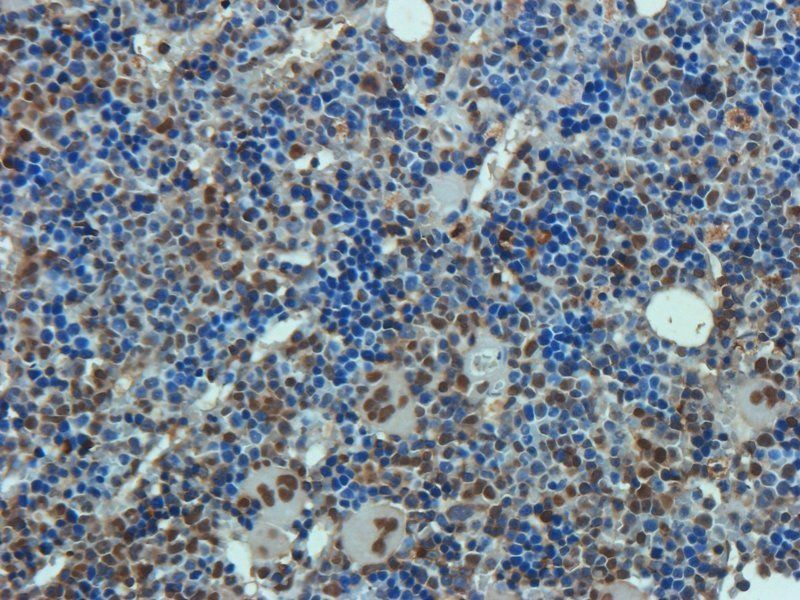
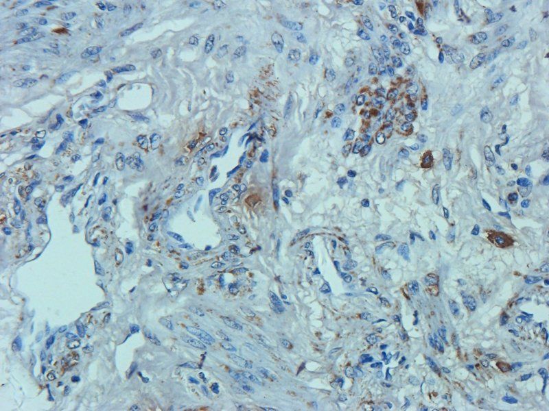
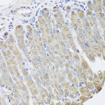
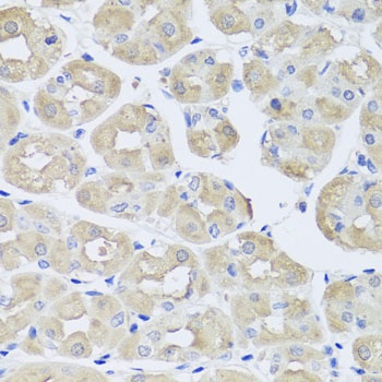
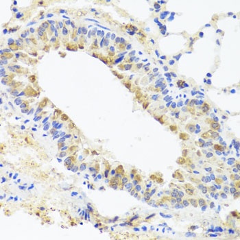
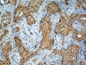
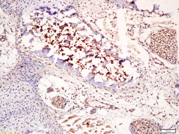
GLA Antibody IHC analysis in formalin fixed and paraffin embedded human Lung carcinoma followed by peroxidase conjugation of the secondary antibody and DAB staining.
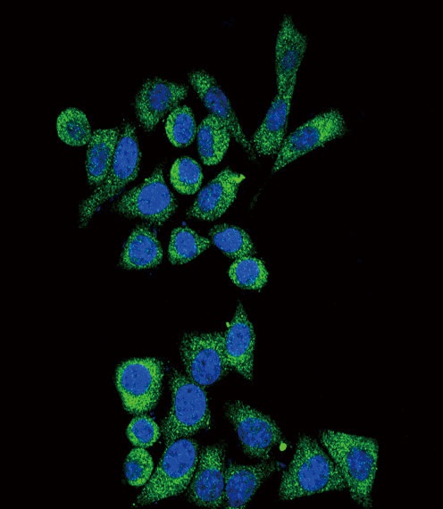
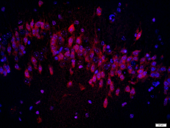
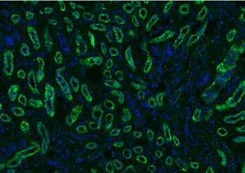
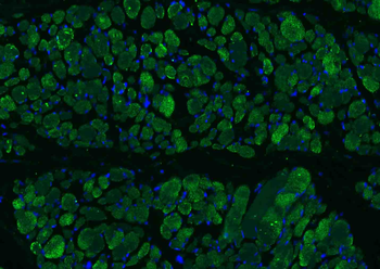
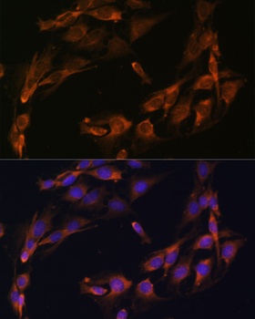
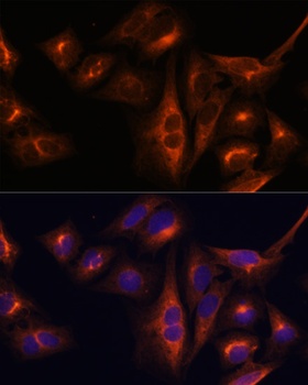
Confocal immunofluorescent analysis of GLA Antibody with Hela cell followed by Alexa Fluor488-conjugated goat anti-rabbit lgG (green). DAPI was used to stain the cell nuclear (blue).
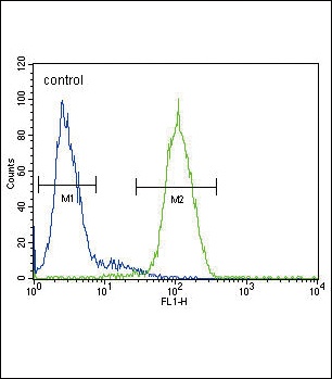
Flow cytometric analysis of HepG2 cells (right histogram) compared to a negative control cell (left histogram). FITC-conjugated goat-anti-rabbit secondary antibodies were used for the analysis.
- Item 1 of 7
Osteocalcin Rabbit Polyclonal Antibody [orb1266]
ELISA, IF, IHC-Fr, IHC-P, WB
Rat
Human, Mouse
Rabbit
Polyclonal
Unconjugated
100 μl, 50 μl - Item 1 of 7
Osteocalcin antibody [orb259644]
ELISA, IHC-P, WB
Human, Mouse, Rat
Rabbit
Polyclonal
Unconjugated
100 μg - Item 1 of 6
- Item 1 of 5
- Item 1 of 2
Osteocalcin Rabbit Polyclonal Antibody [orb10187]
IF, IHC-Fr, IHC-P
Bovine, Canine, Equine, Rat
Human, Mouse
Rabbit
Polyclonal
Unconjugated
50 μl, 100 μl, 200 μl