You have no items in your shopping cart.
Cart summary
Item 1 of 4
Item 1 of 4
GFP antibody (FITC)
Catalog Number: orb345307
| Catalog Number | orb345307 |
|---|---|
| Category | Antibodies |
| Description | GFP antibody (FITC) |
| Species/Host | Goat |
| Clonality | Polyclonal |
| Tested applications | DOT, FLISA, IF, IHC, WB |
| Reactivity | Other |
| Isotype | IgG |
| Immunogen | Recombinant Green Fluorescent Protein (GFP) fusion protein corresponding to the full length amino acid sequence (246aa) derived from the jellyfish Aequorea victoria. |
| Concentration | 1.0 mg/mL |
| Dilution range | FLISA: >1:20,000, IHC: User Optimized, IF: 1:500 - 1:2,500, WB: >1:10,000 |
| Form/Appearance | Lyophilized |
| Purity | GFP Fluorescein Conjugated Antibody was prepared from monospecific antiserum by immunoaffinity chromatography using Green Fluorescent Protein (Aequorea victoria) coupled to agarose beads followed by solid phase adsorption(s) to remove any unwanted reactivities. Assay by immunoelectrophoresis resulted in a single precipitin arc against anti-Goat Serum, anti-Fluorescein and purified and partially purified Green Fluorescent Protein (Aequorea victoria). No reaction was observed against Human, Mouse and Rat Serum Proteins. |
| Conjugation | FITC |
| UniProt ID | P42212 |
| Storage | Store anti-GFP at 4° C prior to restoration. For extended storage aliquot contents and freeze at -20° C or below. Avoid cycles of freezing and thawing. Centrifuge product if not completely clear after standing at room temperature. This product is stable for several weeks at 4° C as an undiluted liquid. Dilute only prior to immediate use. |
| Buffer/Preservatives | 0.01% (w/v) Sodium Azide |
| Alternative names | goat anti-GFP Antibody fluorescein Conjugation, FI Read more... |
| Note | For research use only |
| Application notes | Anti-GFP Fluorescein Conjugated Antibody has been tested by dot blot and western blot and is suitable for immunomicroscopy and flow cytometry or FACS analysis as well as other antibody based fluorescent assays requiring lot-to-lot consistency. |
| Expiration Date | 12 months from date of receipt. |
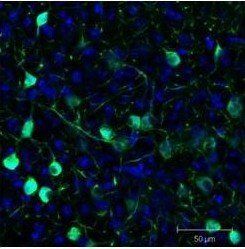
Immunofluorescence analysis of Sf-1:Cre mice crossed to the Z/EG reporter line. Mouse brain (coronal view, 20X magnification) using GFP antibody (FITC)
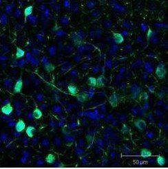
Immunofluorescence analysis of Sf-1:Cre mice crossed to the Z/EG reporter line. Mouse brain (coronal view, 20X magnification) using GFP antibody (FITC)
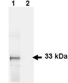
Western blot analysis of Lane 1: HeLa cells. Lane 2: mock transfected HeLa cell lysate. Load: 35 ug per lane using GFP antibody (FITC)
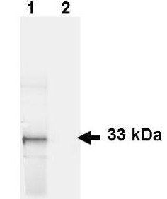
Western blot analysis of Lane 1: HeLa cells. Lane 2: mock transfected HeLa cell lysate. Load: 35 ug per lane using GFP antibody (FITC)
Submit a review
Filter by Rating

- 5 stars

- 4 stars
- 3 stars
- 2 stars
- 1 stars