You have no items in your shopping cart.
Cart summary
Item 1 of 6
Item 1 of 6
FADS2 antibody
Catalog Number: orb1438598
| Catalog Number | orb1438598 |
|---|---|
| Category | Antibodies |
| Description | Rabbit polyclonal antibody to FADS2. |
| Species/Host | Rabbit |
| Clonality | Polyclonal |
| Clone Number | RB31063 |
| Tested applications | FC, IF, IHC-P, WB |
| Reactivity | Human |
| Isotype | Rabbit IgG |
| Immunogen | Synthetic Peptide |
| Dilution range | IF: 1:10~50, WB: 1:1000, WB: 1:1000, WB: 1:1000, IHC-P: 1:10~50, FC: 1:25 |
| Form/Appearance | Purified polyclonal antibody supplied in PBS with 0.09% (W/V) sodium azide. This antibody is purified through a protein A column, followed by peptide affinity purification. |
| Conjugation | Unconjugated |
| MW | 52259 |
| Target | FADS2 |
| UniProt ID | O95864 |
| NCBI | NP_001268430.1, NP_004256.1, NP_001268431.1 |
| Alternative names | Fatty acid desaturase 2, 11419-, Delta(6) fatty ac Read more... |
| Note | For research use only |
| Expiration Date | 12 months from date of receipt. |

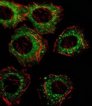
Fluorescent image of A549 cell stained with FADS2 Antibody (N-term) at 1:25 dilution.Secondary antibody:Alexa Fluor® 488 conjugated donkey anti-rabbit antibody(green).Cytoplasmic actin counterstained with Alexa Fluor® 555(red) conjugated Phalloidin.
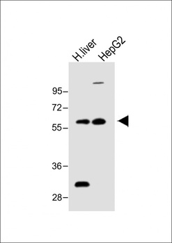
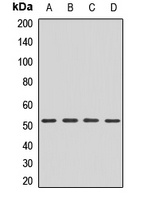
Western blot:All lanes:Anti-FADS2 Antibody (N-term) at 1:1000 dilution.Lane 1:H. liver lysate;Lane 2:HepG2.
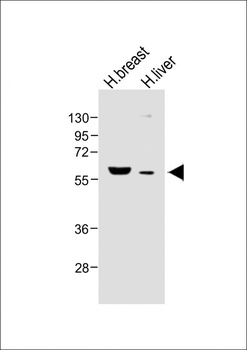
Western blot:All lanes:Anti-FADS2 Antibody (N-term) at 1:1000 dilution.Lane 1:Human breast lysate;Lane 2:Human liver lysate.
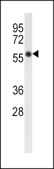
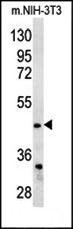
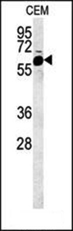
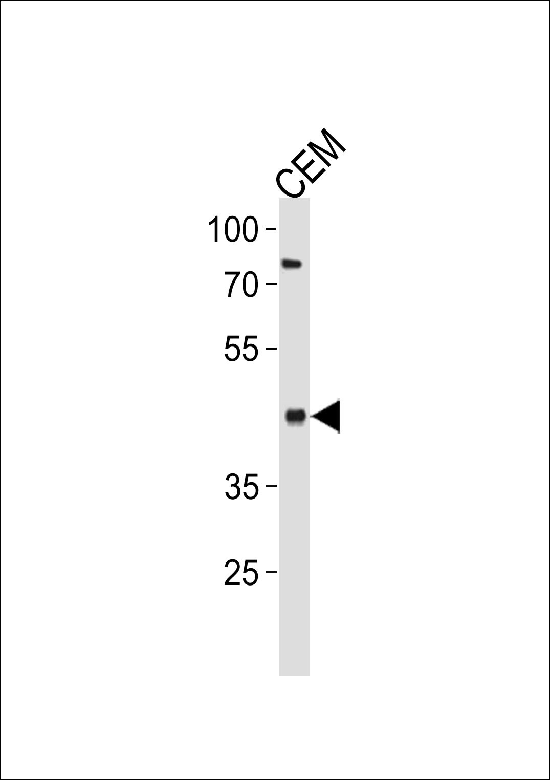
Western blot analysis of HepG2 cell line lysates using FADS2 Antibody (N-term).This demonstrates the FADS2 antibody detected the FADS2 protein (arrow).
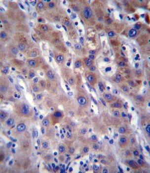
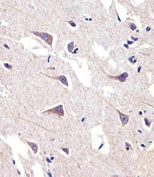
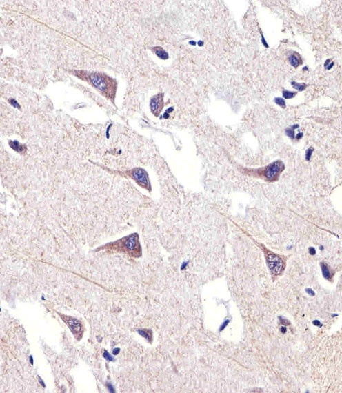
Immunohistochemical analysis of human liver tissue using FADS2 Antibody (N-term), followed by peroxidase conjugation of the secondary antibody and DAB staining.
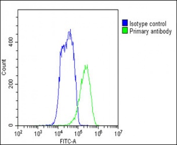
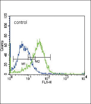
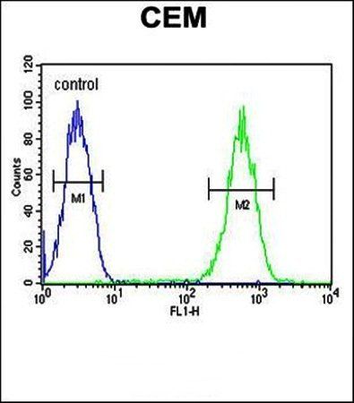
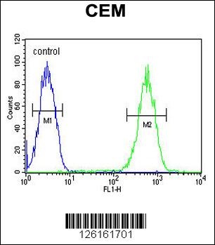
Flow cytometric analysis of HepG2 cells stained with orb1438598(green line) at 1:25 dilution.Secondary antibody was Goat-Anti-Rabbit IgG, DyLight® 488 Conjugated Highly Cross-Adsorbed.Isotype control antibody (blue line) was rabbit IgG1.
- Item 1 of 4
- Item 1 of 4
- Item 1 of 3
- Item 1 of 1


Submit a review
Filter by Rating

- 5 stars

- 4 stars
- 3 stars
- 2 stars
- 1 stars