You have no items in your shopping cart.
Page Not Found
Cart summary
Item 1 of 4
Item 1 of 4
FADS2 Antibody
Catalog Number: orb1271047

| Catalog Number | orb1271047 |
|---|---|
| Category | Antibodies |
| Description | FADS2 Antibody |
| Species/Host | Rabbit |
| Clonality | Polyclonal |
| Tested applications | FC, IF, IHC-P, WB |
| Predicted Reactivity | Monkey |
| Reactivity | Human |
| Isotype | Rabbit Ig |
| Immunogen | This FADS2 antibody is generated from rabbits immunized with a KLH conjugated synthetic peptide between 79-108 amino acids from the N-terminal region of human FADS2. |
| Antibody Type | Primary Antibody |
| Concentration | batch dependent |
| Form/Appearance | Liquid |
| Conjugation | Unconjugated |
| MW | 52 kDa |
| Target | FADS2 |
| UniProt ID | O95864 |
| NCBI | O95864 |
| Storage | Maintain refrigerated at 2-8°C for up to 2 weeks. For long term storage store at -20°C in small aliquots to prevent freeze-thaw cycles. |
| Buffer/Preservatives | Supplied in PBS with 0.09% (W/V) sodium azide. |
| Alternative names | Fatty acid desaturase 2, 11419-, Delta(6) fatty ac Read more... |
| Note | For research use only |
| Application notes | For IF starting dilution is: 1:10~50For WB starting dilution is: 1:1000For IHC-P starting dilution is: 1:10~50For FACS starting dilution is: 1:10~50 |
| Expiration Date | 12 months from date of receipt. |
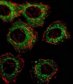
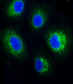
Fluorescent image of A549 cell stained with FADS2 Antibody (N-term). A549 cells were fixed with 4% PFA (20 min), permeabilized with Triton X-100 (0.1%, 10 min), then incubated with FADS2 primary antibody (1:25). For secondary antibody, Alexa Fluor 488 conjugated donkey anti-rabbit antibody (green) was used (1:400). Cytoplasmic actin was counterstained with Alexa Fluor 555 (red) conjugated Phalloidin (7 units/ml). FADS2 immunoreactivity is localized to Cytoplasm and Vesicles significantly.

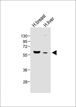
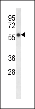
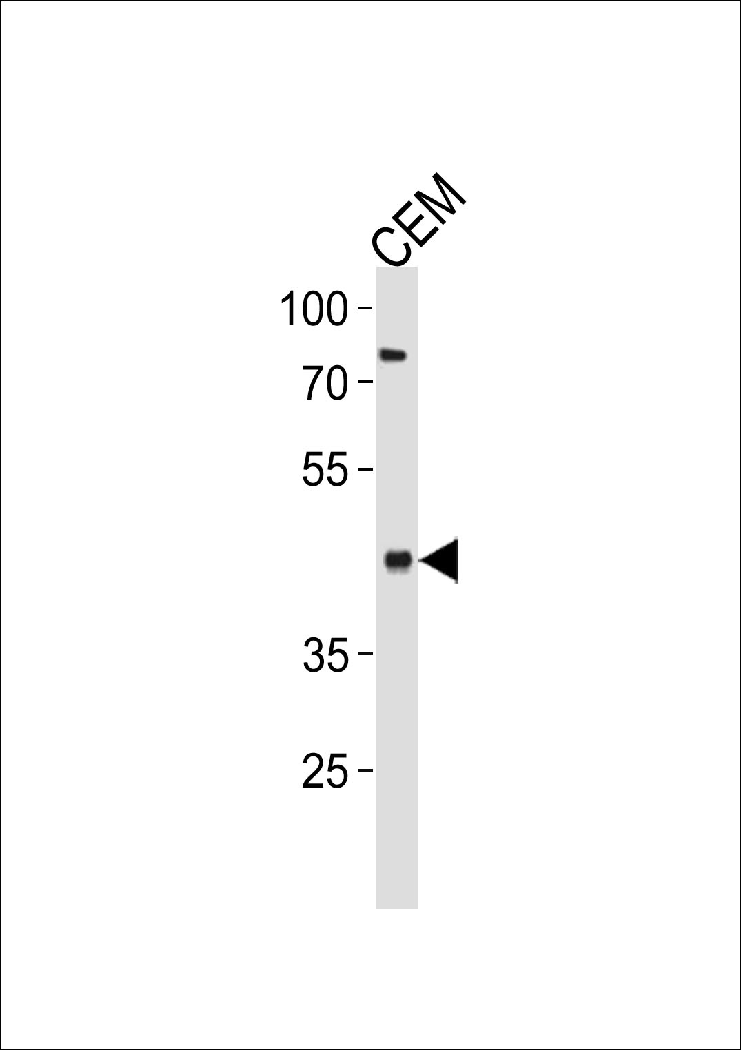
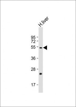
Western blot analysis in HepG2 cell line lysates (35 ug/lane).
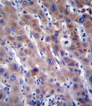
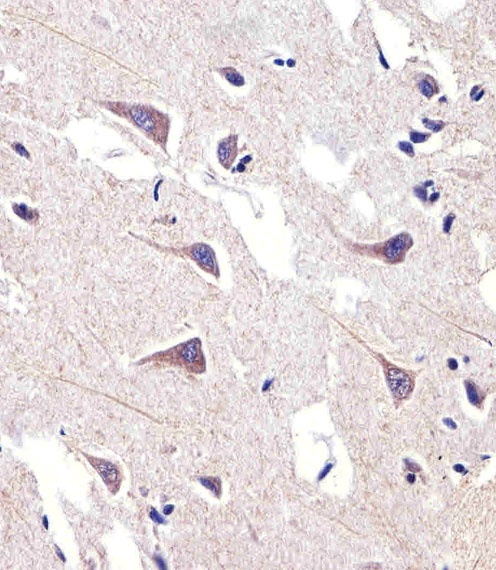
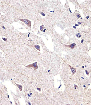
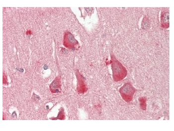
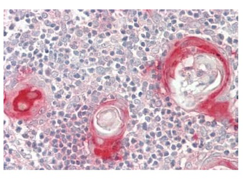
FADS2 Antibody immunohistochemistry analysis in formalin fixed and paraffin embedded human liver tissue followed by peroxidase conjugation of the secondary antibody and DAB staining.
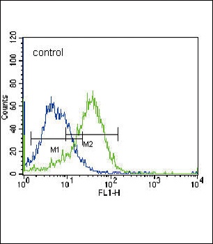
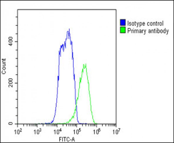
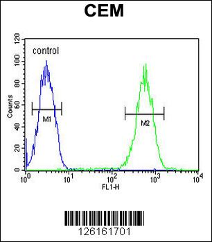
Flow cytometric analysis of K562 cells (right histogram) compared to a negative control cell (left histogram). FITC-conjugated donkey-anti-rabbit secondary antibodies were used for the analysis.
- Item 1 of 6
FADS2 Antibody (N-term) [orb1937594]
FC, IF, IHC-P, WB
Human
Rabbit
Polyclonal
Unconjugated
100 μl, 50 μl - Item 1 of 3
- Item 1 of 3
FADS2 Antibody (Center) [orb1930536]
FC, IHC-P, WB
Other
Human
Rabbit
Polyclonal
Unconjugated
100 μl, 50 μl - Item 1 of 2
- Item 1 of 2