You have no items in your shopping cart.
Cart summary
Item 1 of 3
Item 1 of 3
ERAS Antibody
Catalog Number: orb1268113

| Catalog Number | orb1268113 |
|---|---|
| Category | Antibodies |
| Description | ERAS Antibody |
| Species/Host | Rabbit |
| Clonality | Polyclonal |
| Tested applications | IF, IHC-P, WB |
| Reactivity | Human |
| Isotype | Rabbit Ig |
| Immunogen | This ERAS antibody is generated from rabbits immunized with a KLH conjugated synthetic peptide between 51-81 amino acids from the N-terminal region of human ERAS. |
| Antibody Type | Primary Antibody |
| Concentration | batch dependent |
| Form/Appearance | Liquid |
| Conjugation | Unconjugated |
| MW | 25 kDa |
| Target | ERAS |
| UniProt ID | Q7Z444 |
| NCBI | Q7Z444 |
| Storage | Maintain refrigerated at 2-8°C for up to 2 weeks. For long term storage store at -20°C in small aliquots to prevent freeze-thaw cycles. |
| Buffer/Preservatives | Supplied in PBS with 0.09% (W/V) sodium azide. |
| Alternative names | GTPase ERas, E-Ras, Embryonic stem cell-expressed Read more... |
| Note | For research use only |
| Application notes | For IF starting dilution is: 1:100For WB starting dilution is: 1:1000For IHC-P starting dilution is: 1:10~50 |
| Expiration Date | 12 months from date of receipt. |
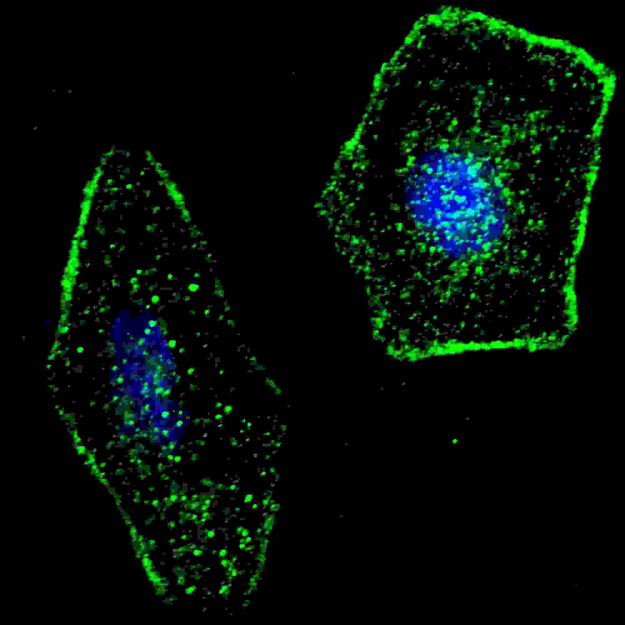
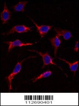
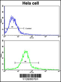
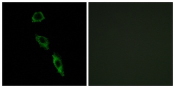
Fluorescent confocal image of SY5Y cells stained with ERAS (F66) antibody. SY5Y cells were fixed with 4% PFA (20 min), permeabilized with Triton X-100 (0.2%, 30 min). Cells were then incubated with ERAS (F66) primary antibody (1:100, 2 h at room temperature). For secondary antibody, Alexa Fluor 488 conjugated donkey anti-rabbit antibody (green) was used (1:1000, 1h). Nuclei were counterstained with Hoechst 33342 (blue) (10 ug/ml, 5 min). ERAS immunoreactivity is localized to the plasma membrane of SY5Y cells.

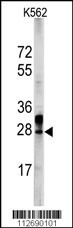
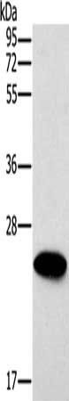
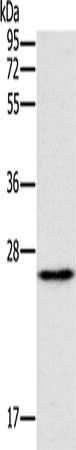
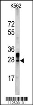
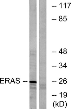
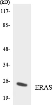
Western blot analysis of ERAS Antibody (F66) in HL60 cell line lysates (35 ug/lane)
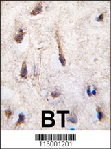
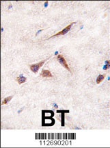
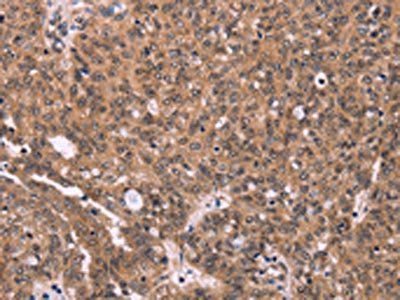
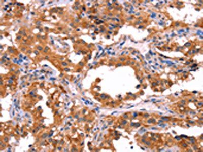
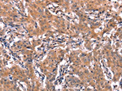
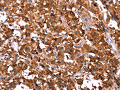
Formalin-fixed and paraffin-embedded human brain tissue reacted with ERAS antibody (F66), which was peroxidase-conjugated to the secondary antibody, followed by DAB staining.
- Item 1 of 4
- Item 1 of 3
- Item 1 of 3
- Item 1 of 4
ERAS Antibody (N-term) [orb1935563]
FC, IF, IHC-P, WB
Human
Rabbit
Polyclonal
Unconjugated
100 μl, 50 μl - Item 1 of 3