You have no items in your shopping cart.
Cart summary
Item 1 of 7
Item 1 of 7
EpCAM Antibody
Catalog Number: orb749657

| Catalog Number | orb749657 |
|---|---|
| Category | Antibodies |
| Description | EpCAM may act as a physical homophilic interaction molecule between intestinal epithelial cells (IECs) and intraepithelial lymphocytes (IELs) at the mucosal epithelium for providing immunological barrier as a first line of defense against mucosal infection. [UniProt] |
| Clonality | Monoclonal |
| Species/Host | Mouse |
| Isotype | Mouse IgG1, kappa |
| Conjugation | Unconjugated |
| Reactivity | Human |
| Immunogen | Recombinant human protein was used as the immunogen for the EpCAM antibody. |
| UniProt ID | P16422 |
| Tested applications | FACS, IF, IHC-P, WB |
| Dilution range | Flow cytometry: 1-2ug/million cells,Immunofluorescence: 1-2ug/ml,Western blot: 1-2ug/ml,Immunohistochemistry (FFPE): 1-2ug/ml for 30 min at RT |
| Application notes | Optimal dilution of the EpCAM antibody should be determined by the researcher.1. Staining of formalin-fixed tissues requires boiling tissue sections in 10mM Citrate buffer, pH 6.0, for 10-20 min followed by cooling at RT for 20 min2. The prediluted format is supplied in a dropper bottle and is optimized for use in IHC. After epitope retrieval step (if required), drip mAb solution onto the tissue section and incubate at RT for 30 min. |
| Antibody Type | Primary Antibody |
| Clone Number | EGP40/837 |
| Formula | 0.2 mg/ml in 1X PBS with 0.1 mg/ml BSA (US sourced) and 0.05% sodium azide |
| Storage | Maintain refrigerated at 2-8°C for up to 2 weeks. For long term storage store at -20°C in small aliquots to prevent freeze-thaw cycles. |
| Hazard Information | This EpCAM antibody is available for research use only. |
| Note | For research use only |
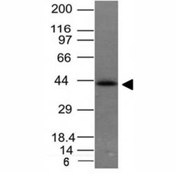
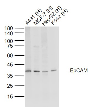
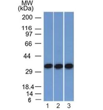
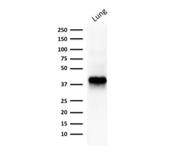
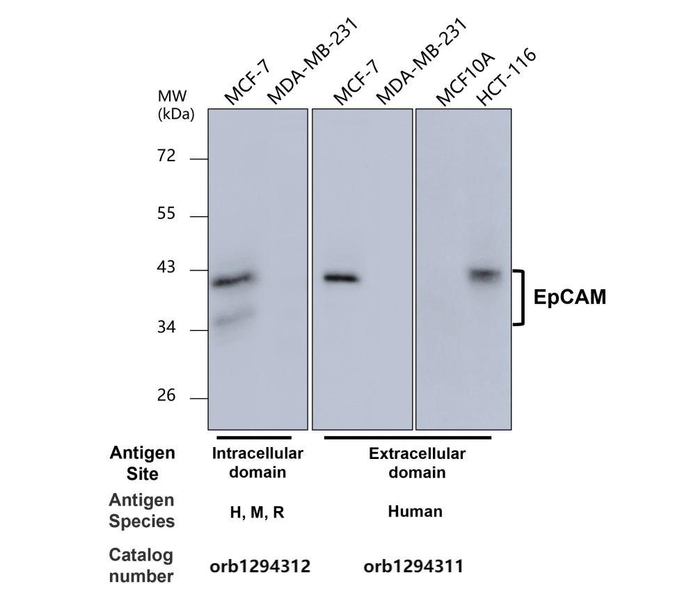
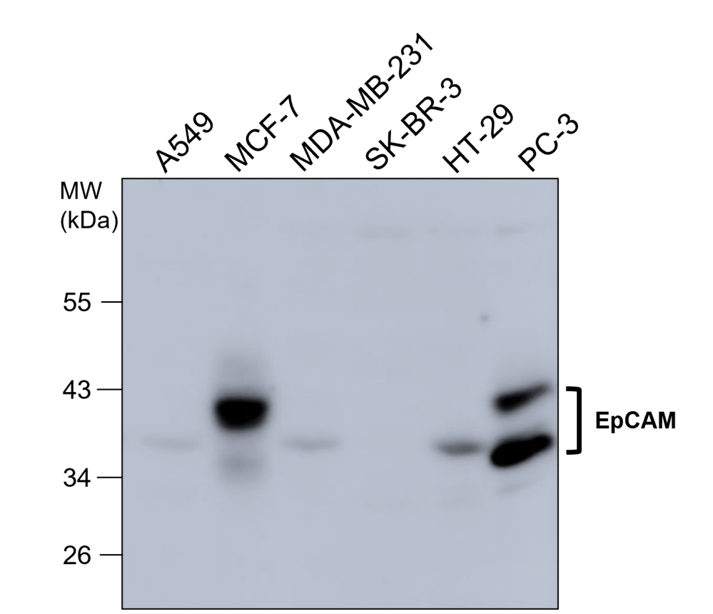
Western blot of HCT116 cell lysate using EpCAM antibody (EGP40/837). Expected molecular weight: ~35 kDa (unmodified), 40-43 kDa (glycosylated).
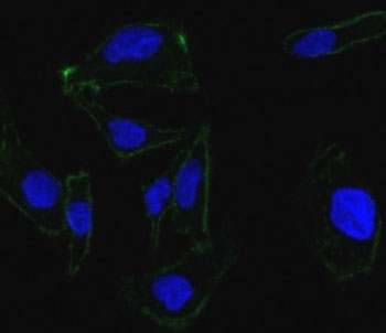
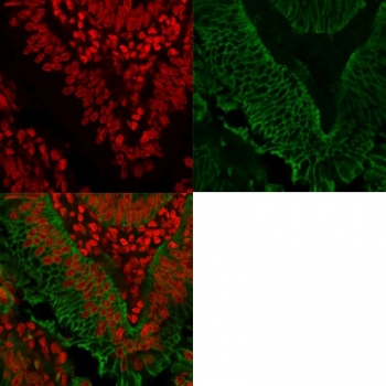
Confocal Immunofluorescent analysis of SK-OV-3 cells using AF488-labeled EpCAM antibody (green). DAPI was used to stain the cell nuclei (blue).
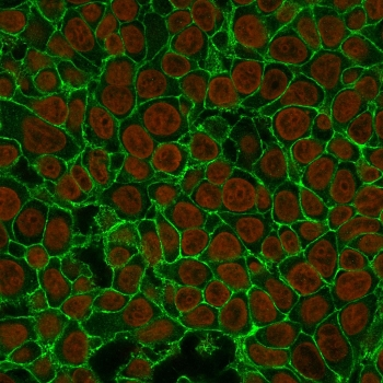
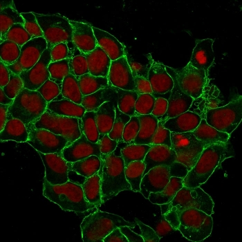
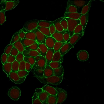
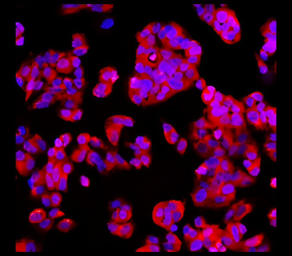
Immunofluorescent staining of human MCF7 cells with EpCAM antibody (clone EGP40/837, green) and Reddot nuclear stain (red).
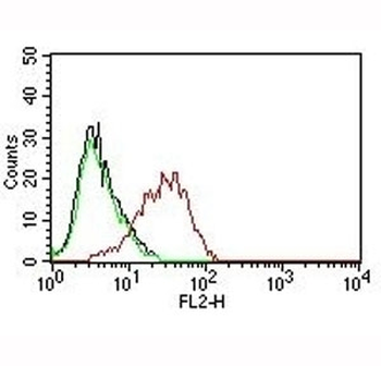
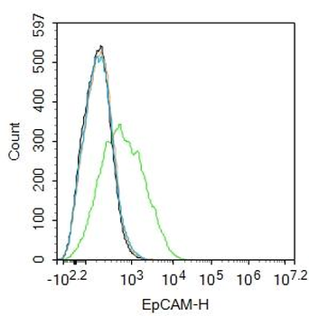
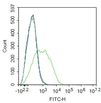
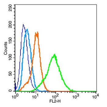
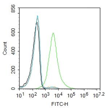
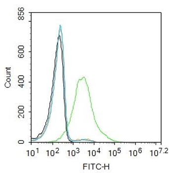
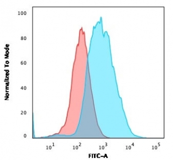
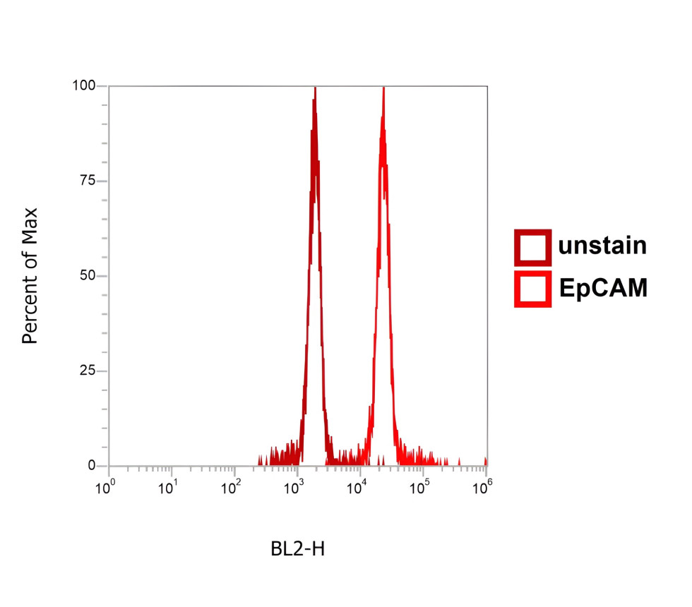
Flow cytometry testing of human MCF-7 cells. Black: cells alone; Green: isotype control; Red: PE-labeled EpCAM antibody.
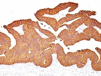
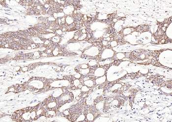
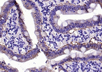
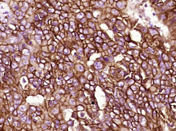
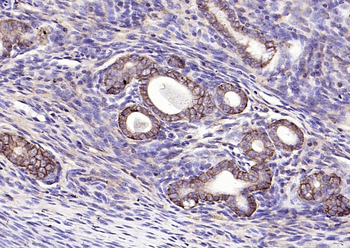
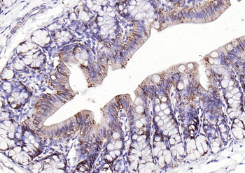
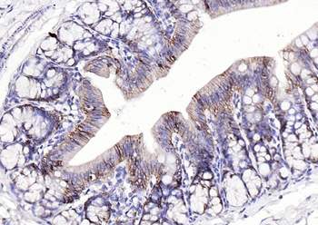
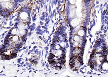
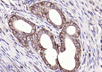
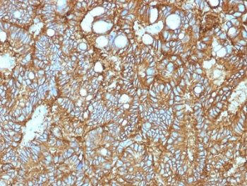
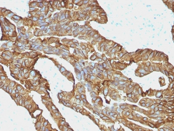
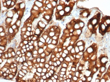
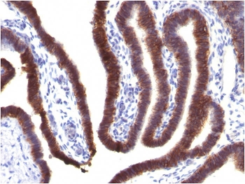
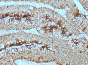
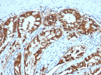
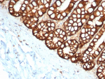
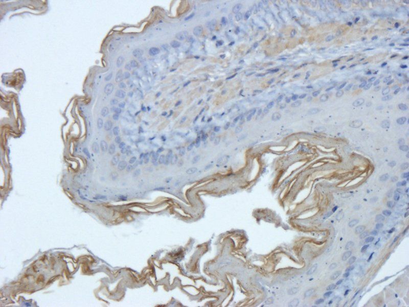
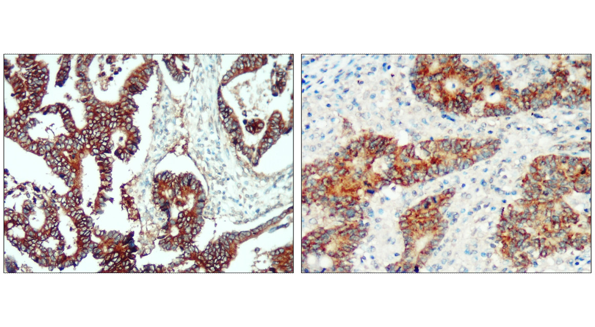
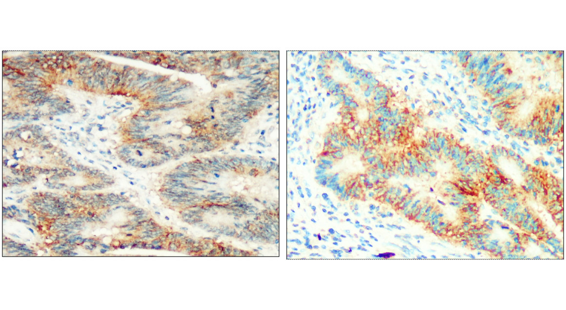
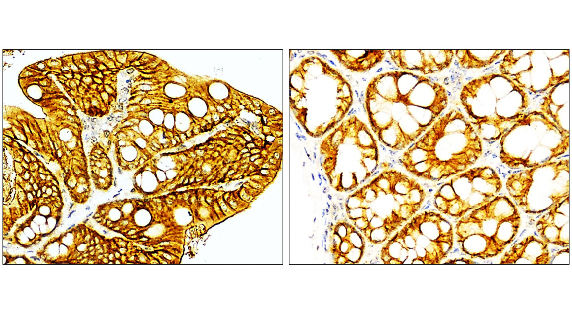
IHC: Formalin-fixed, paraffin-embedded human colon carcinoma stained with EpCAM antibody.

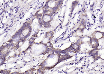
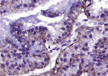
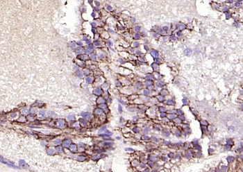
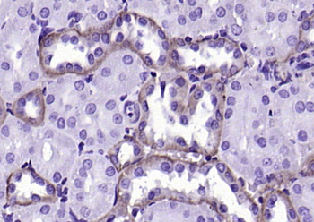
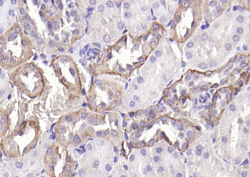
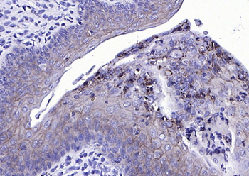
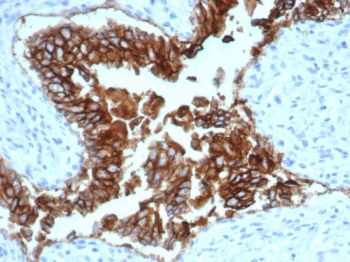
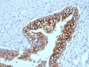
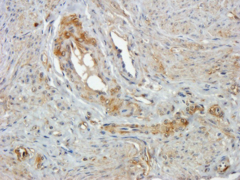
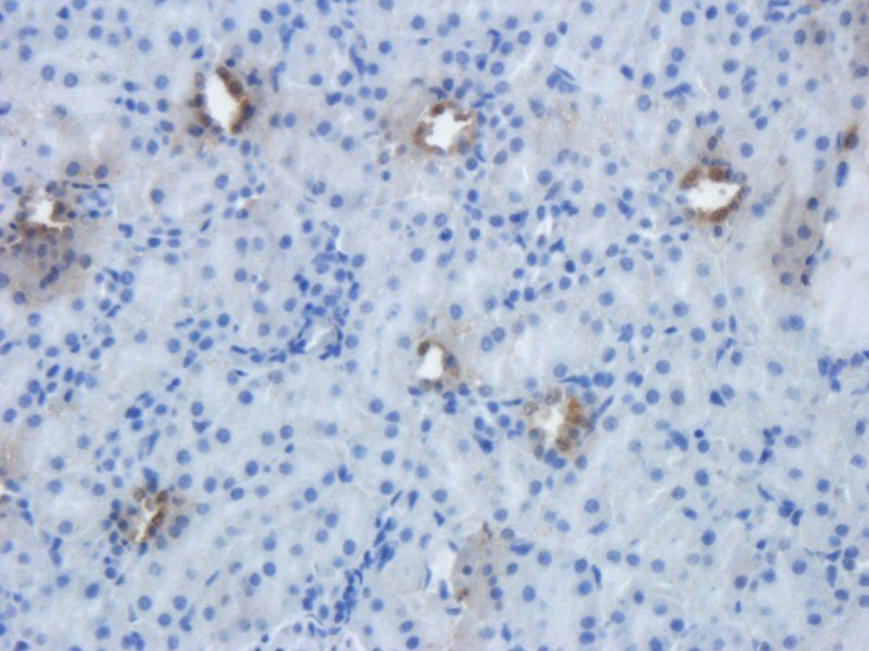
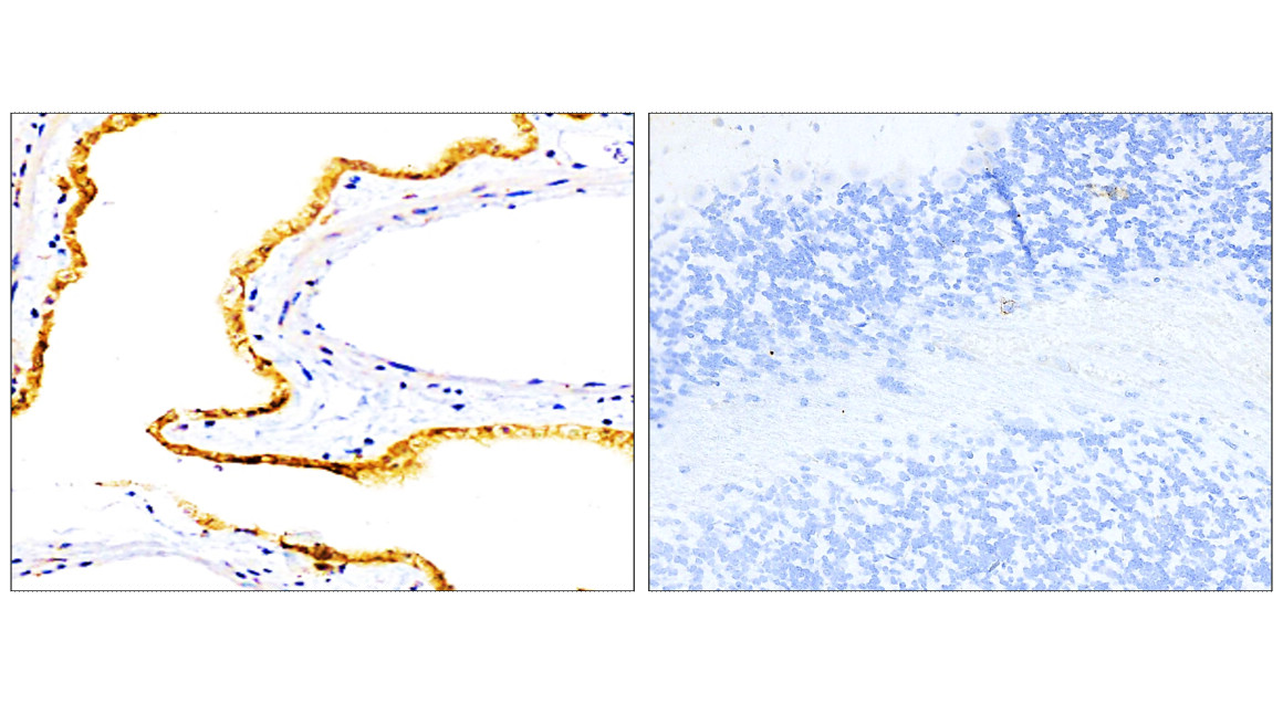
IHC: Formalin-fixed, paraffin-embedded human ovarian carcinoma stained with EpCAM antibody.
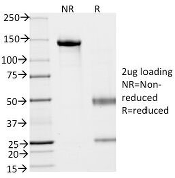
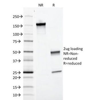
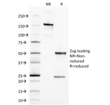
SDS-PAGE analysis of purified, BSA-free EpCAM antibody (clone EGP40/837) as confirmation of integrity and purity.
- Item 1 of 20
EpCAM Rabbit Polyclonal Antibody [orb10183]
FC, IF, IHC-Fr, IHC-P, WB
Rat
Human, Mouse, Rat
Rabbit
Polyclonal
Unconjugated
100 μl, 50 μl, 200 μl - Item 1 of 11
EpCAM Antibody / Extracellular domain [orb606357]
FACS, IF, IHC-P, WB
Canine, Feline, Human
Mouse
Monoclonal
Unconjugated
20 μg, 100 μg - Item 1 of 9
EpCAM Antibody [orb639787]
IF, IHC-P, WB
Canine, Feline, Human
Mouse
Monoclonal
Unconjugated
20 μg, 100 μg - Item 1 of 5
- Item 1 of 8
EpCAM/CD326 (Extracellular domain) Antibody [orb1294311]
FC, IF, IHC, WB
Human, Mouse
Rabbit
Polyclonal
Unconjugated
25 μl, 100 μl