You have no items in your shopping cart.
Cart summary
Item 1 of 4
Item 1 of 4
EPCAM Antibody
Catalog Number: orb1671668

| Catalog Number | orb1671668 |
|---|---|
| Category | Antibodies |
| Description | EPCAM Antibody |
| Clonality | Recombinant |
| Clone Number | HEA125 |
| Tested applications | FC, IF, IHC, IP, RIA, WB |
| Reactivity | Human |
| Isotype | IgG1 kappa |
| Immunogen | This antibody was raised by immunising BALB/c mice with the human colon carcinoma cell line HT-29. |
| Concentration | batch dependent |
| Conjugation | Unconjugated |
| Target | EPCAM |
| UniProt ID | P16422 |
| Storage | Store at 4°C for up to 3 months. For longer storage, aliquot and store at -20°C. |
| Buffer/Preservatives | PBS with 0.02% Proclin 300. |
| Alternative names | CD326, Ep-CAM, Epithelial cell adhesion molecule, Read more... |
| Note | For research use only |
| Expiration Date | 12 months from date of receipt. |
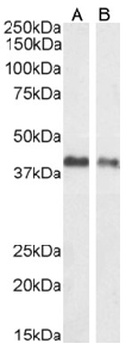
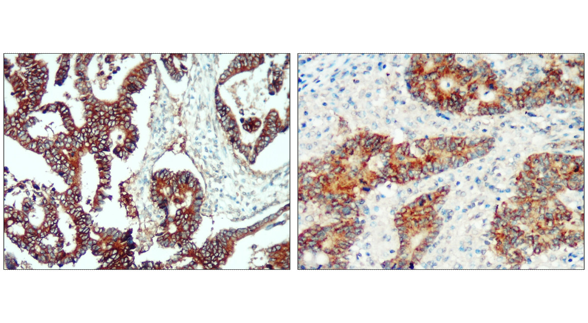
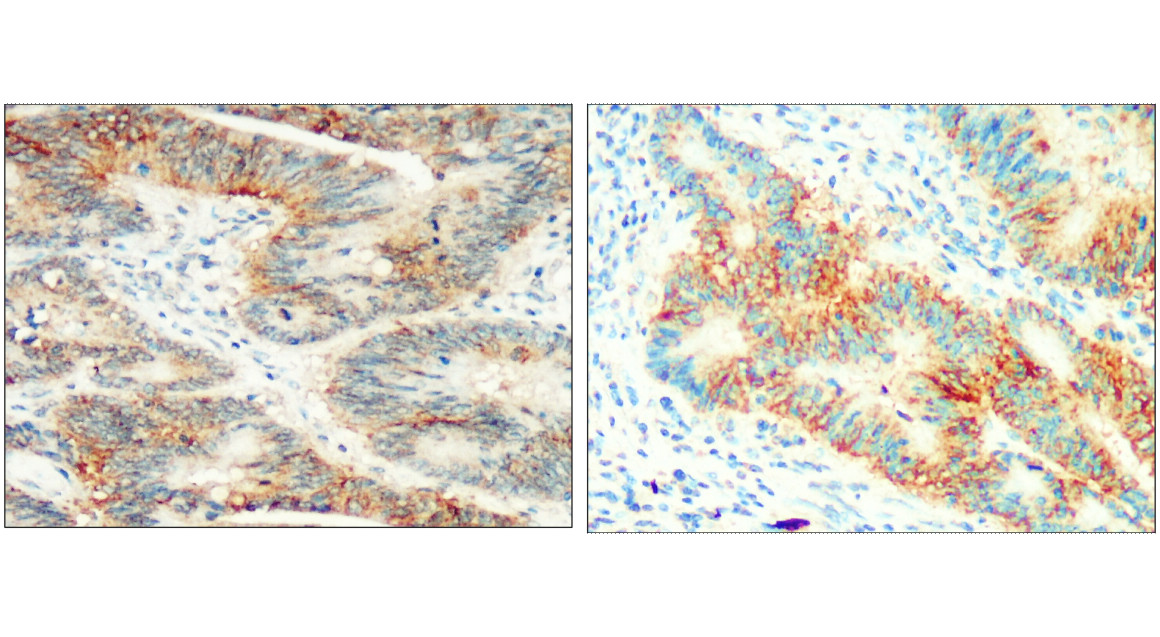
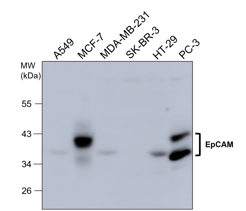
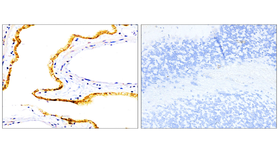
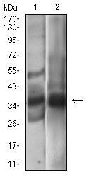
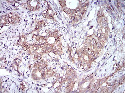
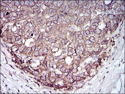
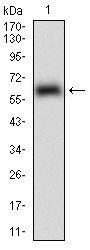
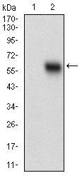
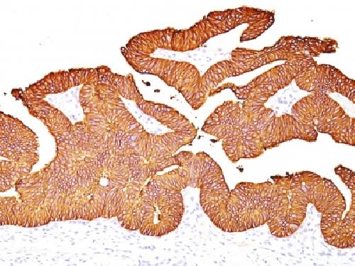
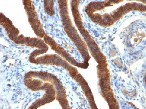
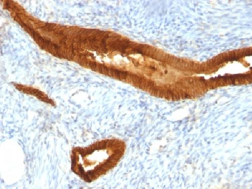
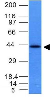
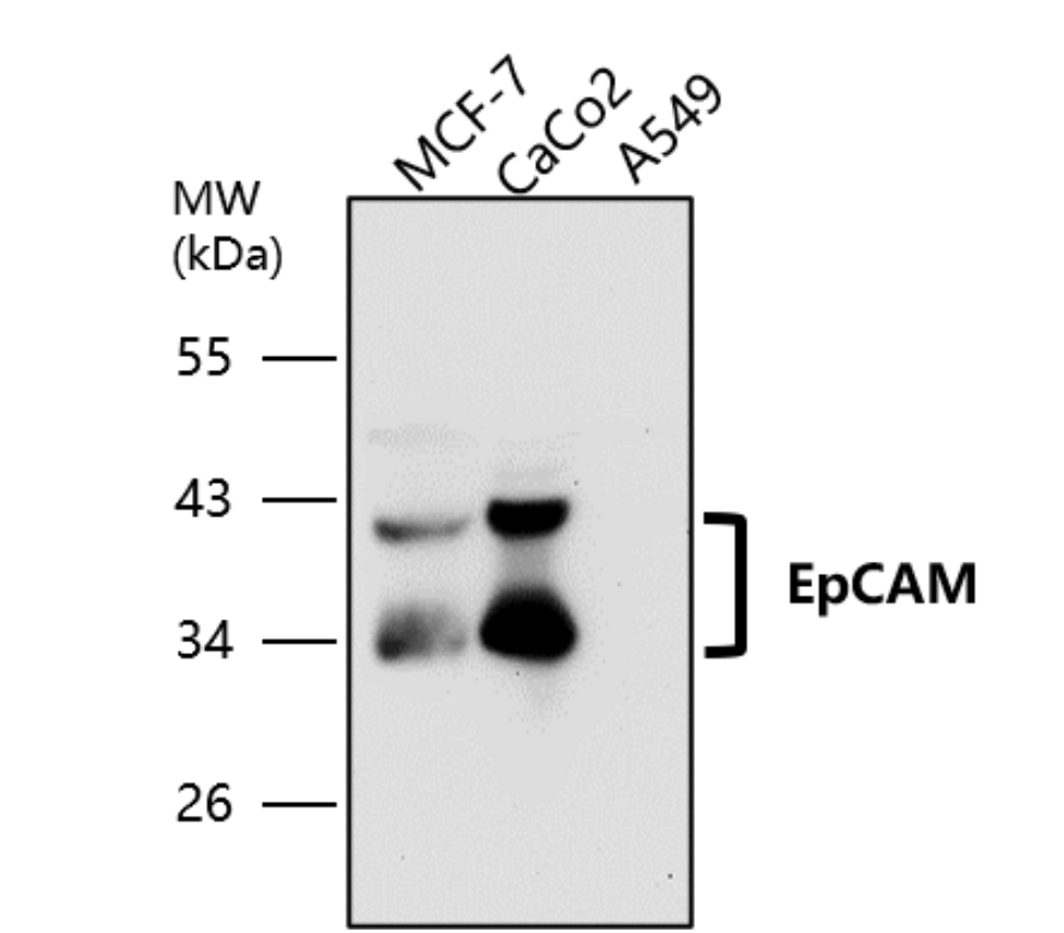
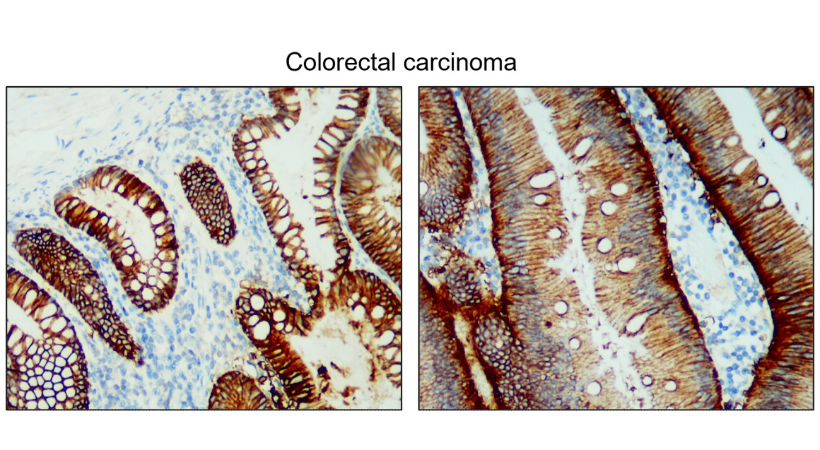
Western Blot using anti-EpCAM antibody HEA125, human breast carcinoma(A) and human colorectal carcinoma(B) tissue lysate (35 ug protein in RIPA buffer) was resolved on an SDS PAGE gel and blots probed with the chimeric mouse IgG version of HEA125 (orb1671668) at 1 ug/ml before detection using an anti-mouse secondary antibody. A primary incubation of 1h was used and protein was detected by chemiluminescence.
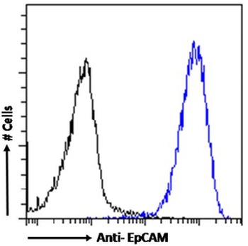
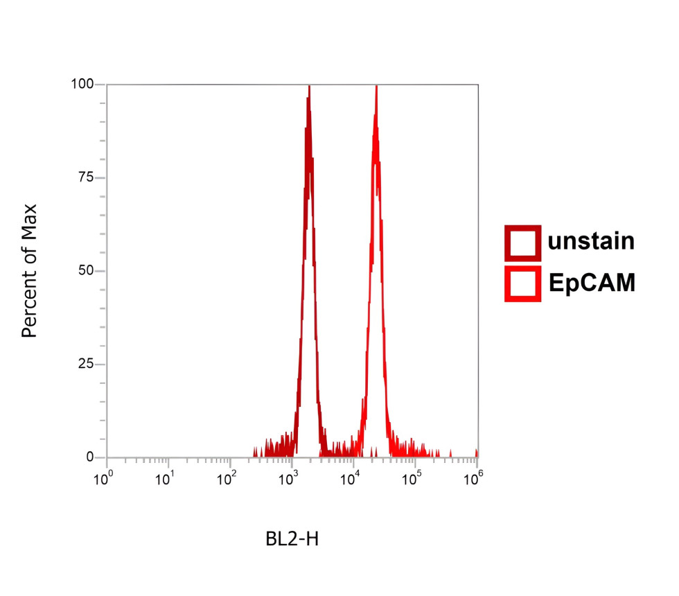
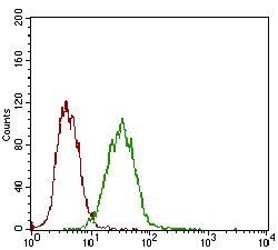
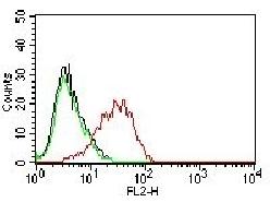
Flow-cytometry using the anti-EpCAM antibody HEA125. Caco-2 cells were stained with anti-unknown IgG antibody (.1; isotype control - black line) or the mouse IgG-chimeric version of HEA125 (orb1671668 - blue line) at a dilution of 1:100 for 1h at RT. After washing- bound antibody was detected using a goat anti-mouse IgG AlexaFluor® 488 antibody at a dilution of 1:1000 and cells analyzed using a FACSCanto flow-cytometer.
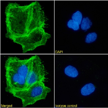
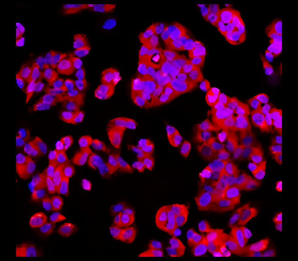
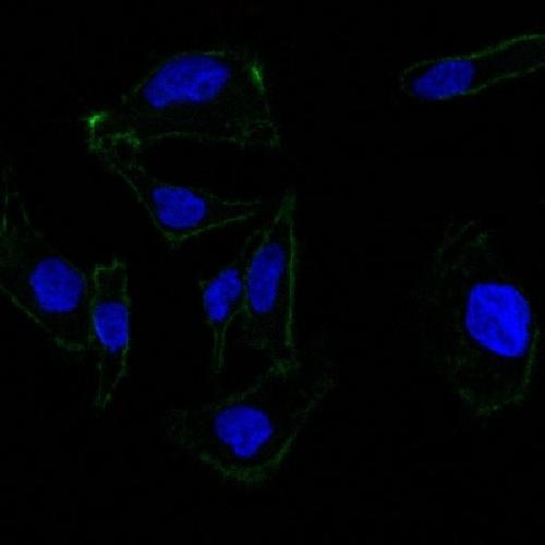
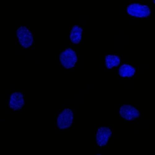
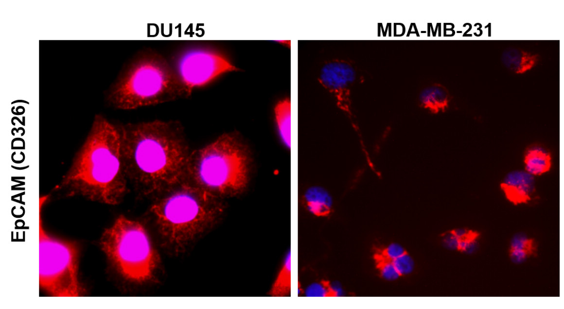
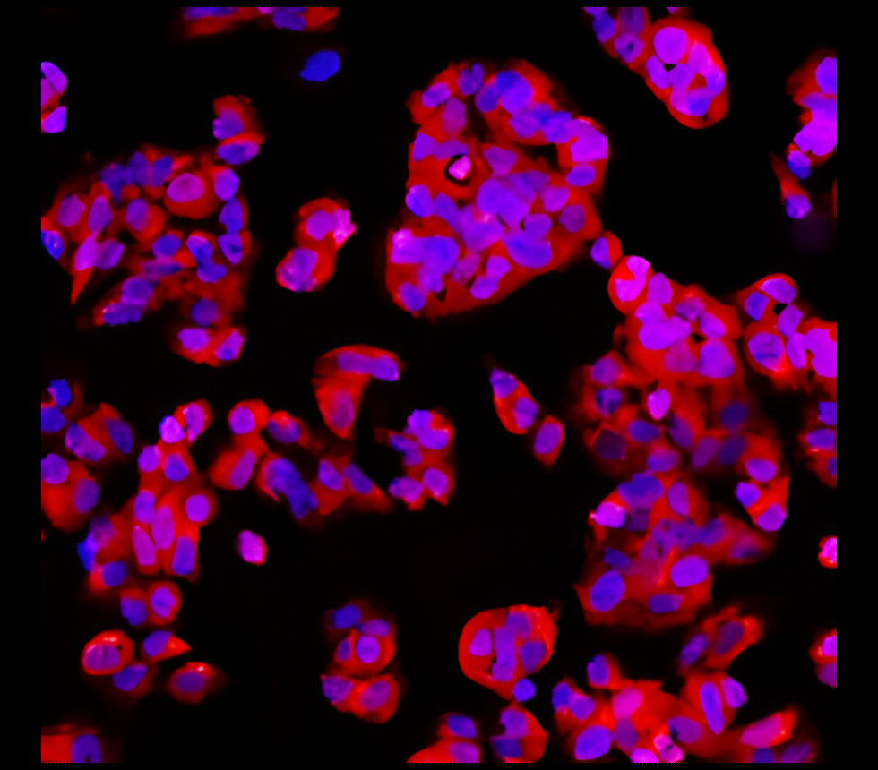
Immunofluorescence staining of Caco-2 cells using anti-EpCAM (HEA125). Immunofluorescence analysis of paraformaldehyde fixed Caco-2 cells stained with the chimeric mouse IgG version of HEA125 (orb1671668) at 10 ug/ml followed by Alexa Fluor® 488 secondary antibody (2 ug/ml)- showing membrane staining. The nuclear stain is DAPI (blue). Panels show from left-right- top-bottom. 1- DAPI- merged channels and an isotype control. The isotype control was stained with anti-unknown antibody (.1) followed by Alexa Fluor® 488 secondary antibody.
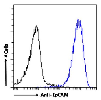
Flow cytometry using the Anti-EpCAM antibody HEA125. Caco-2 cells were stained with anti-unknown specificity antibody (.1; isotype control - black line) or the mouse IgG1 version of HEA125 (orb1671668 - blue line) at a dilution of 1:100 for 1h at RT. After washing- the bound antibody was detected using a goat anti-mouse IgG AlexaFluor® 488 antibody at a dilution of 1:1000 and cells analyzed using a FACSCanto flow-cytometer.
- Item 1 of 5
- Item 1 of 8
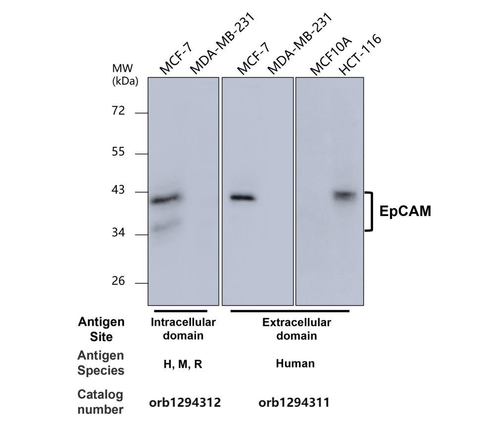
EpCAM (Extracellular domain) antibody [orb1294311]
IF, IHC, WB
Human
Rabbit
Polyclonal
Unconjugated
100 μl, 25 μl - Item 1 of 7
- Item 1 of 7
EpCAM antibody [orb389162]
FC, IF, IHC-P, WB
Canine, Feline, Human
Mouse
Monoclonal
Unconjugated
100 μg, 20 μg - Item 1 of 7
EpCAM (intracellular domain) antibody [orb1294312]
IF, IHC, WB
Human, Mouse, Rabbit
Rabbit
Polyclonal
Unconjugated
100 μl, 25 μl
Submit a review
Filter by Rating

- 5 stars

- 4 stars
- 3 stars
- 2 stars
- 1 stars
