You have no items in your shopping cart.
Cart summary
Item 1 of 7
Item 1 of 7
DDIT3 Antibody
Catalog Number: orb1269882

| Catalog Number | orb1269882 |
|---|---|
| Category | Antibodies |
| Description | DDIT3 Antibody |
| Target | DDIT3 |
| Clonality | Polyclonal |
| Isotype | Rabbit Ig |
| Conjugation | Unconjugated |
| Reactivity | Human, Mouse |
| Predicted Reactivity | Bovine, Hamster |
| Form/Appearance | Liquid |
| Concentration | batch dependent |
| Buffer/Preservatives | Supplied in PBS with 0.09% (W/V) sodium azide. |
| Purification | This antibody is purified through a protein A column, followed by peptide affinity purification. |
| Immunogen | This DDIT3 antibody is generated from rabbits immunized with a KLH conjugated synthetic peptide between 120-149 amino acids from the C-terminal region of human DDIT3. |
| UniProt ID | P35638 |
| MW | 19 kDa |
| Tested applications | FC, IF, WB |
| Application notes | For FACS starting dilution is: 1:25For WB starting dilution is: 1:2000For IF starting dilution is: 1:10~50 |
| Antibody Type | Primary Antibody |
| Storage | Maintain refrigerated at 2-8°C for up to 2 weeks. For long term storage store at -20°C in small aliquots to prevent freeze-thaw cycles. |
| Alternative names | DNA damage-inducible transcript 3 protein, DDIT-3, Read more... |
| Note | For research use only |
| NCBI | P35638 |
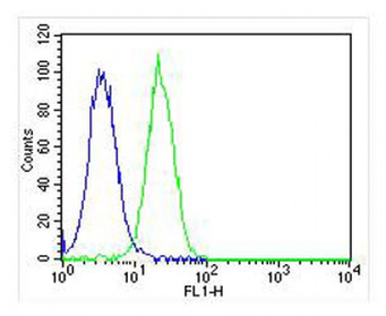
Overlay histogram showing Hela cells stained with Antibody (green line). The cells were fixed with 2% paraformaldehyde (10 min) and then permeabilized with 90% methanol for 10 min. The cells were then icubated in 2% bovine serum albumin to block non-specific protein-protein interactions followed by the antibody (1:25 dilution) for 60 min at 37oC. The secondary antibody used was Goat-Anti-Rabbit IgG, DyLight 488 Conjugated Highly Cross-Adsorbed (OH191631) at 1/400 dilution for 40 min at 37oC. Isotype control antibody (blue line) was rabbit IgG (1 ug/1x10^6 cells) used under the same conditions. Acquisition of >10000 events was performed.
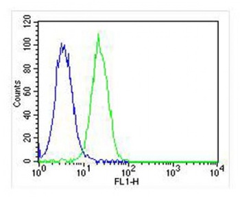
Overlay histogram showing Hela cells stained with Antibody (green line). The cells were fixed with 2% paraformaldehyde (10 min) and then permeabilized with 90% methanol for 10 min. The cells were then icubated in 2% bovine serum albumin to block non-specific protein-protein interactions followed by the antibody (1:25 dilution) for 60 min at 37oC. The secondary antibody used was Goat-Anti-Rabbit IgG, DyLight 488 Conjugated Highly Cross-Adsorbed (OH191631) at 1/400 dilution for 40 min at 37oC. Isotype control antibody (blue line) was rabbit IgG (1 ug/1x10^6 cells) used under the same conditions. Acquisition of >10000 events was performed.
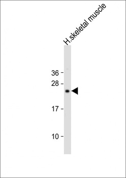
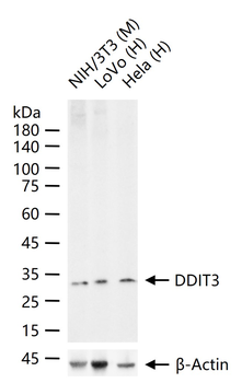
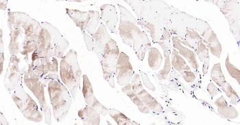
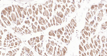
Western Blot at 1:2000 dilution + human skeletal muscle lysate Lysates/proteins at 20 ug per lane.
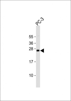
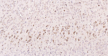
Western Blot at 1:2000 dilution + PC-3 whole cell lysate Lysates/proteins at 20 ug per lane.
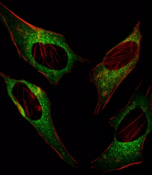
Fluorescent image of Hela cell stained with.Hela cells were fixed with 4% PFA (20 min), permeabilized with Triton X-100 (0.1%, 10 min), then incubated with DDIT3 primary antibody (1:25). For secondary antibody, Alexa Fluor 488 conjugated donkey anti-rabbit antibody (green) was used (1:400). Cytoplasmic actin was counterstained with Alexa Fluor 555 (red) conjugated Phalloidin (7 units/ml). DDIT3 immunoreactivity is localized to Cytoplasm significantly.
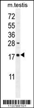
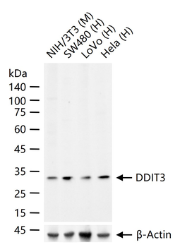
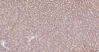
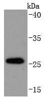
Western blot analysis in mouse testis tissue lysates (35 ug/lane).This demonstrates the DDIT3 antibody detected the DDIT3 protein (arrow).
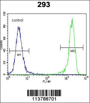
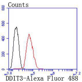
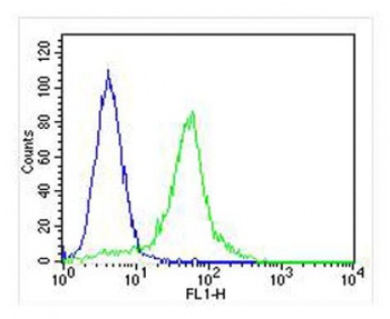
Flow cytometric analysis of 293 cells (right histogram) compared to a negative control cell (left histogram). FITC-conjugated goat-anti-rabbit secondary antibodies were used for the analysis.
- Item 1 of 16
DDIT3 Recombinant Rabbit Monoclonal Antibody [orb2563499]
IF, IHC-Fr, IHC-P, WB
Mouse, Rat
Human, Mouse, Rat
Rabbit
Recombinant
Unconjugated
25 μl, 50 μl, 100 μl - Item 1 of 11
DDIT3 Rabbit Polyclonal Antibody [orb500776]
IF, IHC-Fr, IHC-P, WB
Bovine
Human, Mouse, Rat
Rabbit
Polyclonal
Unconjugated
100 μl, 200 μl, 50 μl - Item 1 of 6
DDIT3 Rabbit Polyclonal Antibody [orb10684]
IF, IHC-Fr, IHC-P
Mouse, Rat
Human, Mouse, Rat
Rabbit
Polyclonal
Unconjugated
50 μl, 100 μl, 200 μl - Item 1 of 7
- Item 1 of 7
DDIT3 Antibody (C-term A135) [orb1937840]
FC, IF, WB
Hamster
Human, Mouse
Rabbit
Polyclonal
Unconjugated
100 μl, 50 μl