You have no items in your shopping cart.
Cart summary
Item 1 of 11
Item 1 of 11
DDB1 Antibody
Catalog Number: orb312104
| Catalog Number | orb312104 |
|---|---|
| Category | Antibodies |
| Description | DDB1 Antibody |
| Species/Host | Rabbit |
| Clonality | Polyclonal |
| Tested applications | FC, ICC, IF, IHC, IHC-Fr, WB |
| Predicted Reactivity | Bovine, Canine, Equine, Hamster, Monkey, Rabbit |
| Reactivity | Human, Mouse, Rat |
| Isotype | Rabbit IgG |
| Immunogen | E.coli-derived human DDB1 recombinant protein (Position: S1011-H1140). Human DDB1 shares 99.2% amino acid (aa) sequence identity with both mouse and rat DDB1. |
| Concentration | Adding 0.2 ml of distilled water will yield a concentration of 500 μg/ml. |
| Dilution range | Western blot, 0.1-0.5μg/ml Immunohistochemistry (Paraffin-embedded Section), 0.5-1μg/ml, By Heat Immunohistochemistry (Frozen Section), 0.5-1μg/ml Immunocytochemistry, 0.5-1μg/ml Immunocytochemistry/Immunofluorescence, 2μg/ml Flow Cytometry, 1-3μg/1x106 cells |
| Form/Appearance | Lyophilized |
| Conjugation | Unconjugated |
| MW | 126968 MW |
| UniProt ID | Q16531 |
| Storage | Store at -20˚C for one year from date of receipt. After reconstitution, at 4˚C for one month. It can also be aliquotted and stored frozen at -20˚C for six months. Avoid repeated freeze-thaw cycles. |
| Alternative names | DNA damage-binding protein 1;DDB p127 subunit;DNA Read more... |
| Note | For research use only |
| Application notes | Tested Species: In-house tested species with positive results. By Heat: Boiling the paraffin sections in 10mM citrate buffer, pH6.0, for 20mins is required for the staining of formalin/paraffin sections. Other applications have not been tested. Optimal dilutions should be determined by end users. . Add 0.2ml of distilled water will yield a concentration of 500ug/ml. |
| Expiration Date | 12 months from date of receipt. |
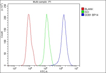
Flow Cytometry analysis of 293T cells using anti-DDB1 antibody (Blue line).Isotype control antibody (Green line) was rabbit IgG .Unlabelled sample (Red line) was also used as a control.
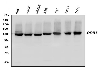
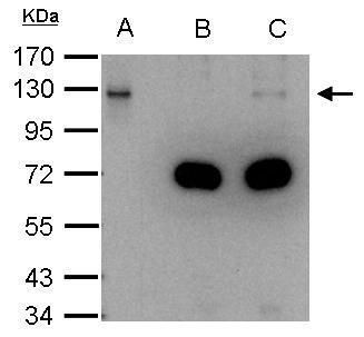
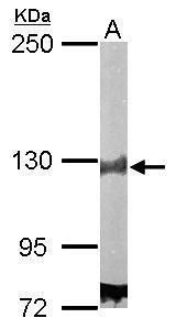
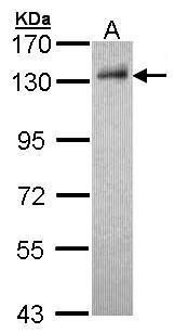
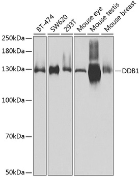
WB analysis of DDB1 using anti-DDB1 antibody.Lane 1:human HeLa cell; 2:human HepG2 cell; 3:human HEK293 cell; 4:human K562 cell; 5:human Raji cell; 6:human CACO-2 cell; 7:human THP-1 cell.
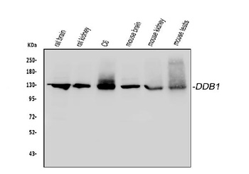
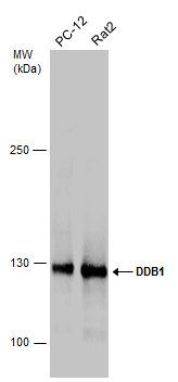
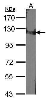
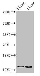
WB analysis of DDB1 using anti-DDB1 antibody.Lane 1:rat brain tissue; 2:rat kidney tissue; 3:rat C6 cell; 4:mouse brain tissue; 5:mouse kidney tissue; 6:mouse testis tissue.
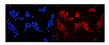
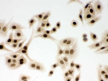
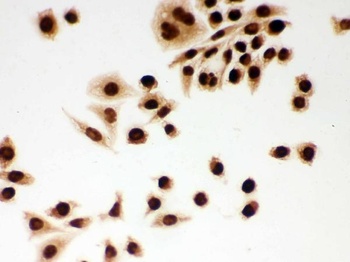
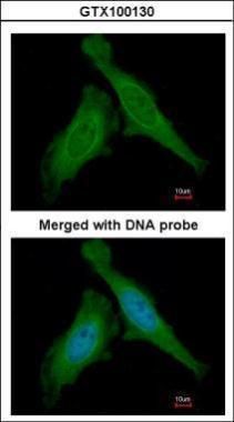
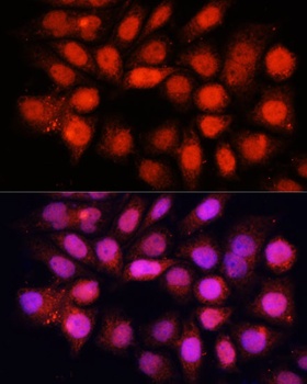
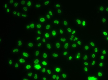
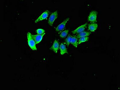
IF analysis of DDB1 using anti-DDB1 antibody. DDB1 was detected in immunocytochemical section of A431 cells.
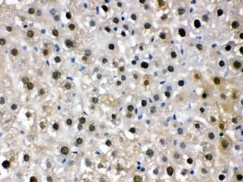
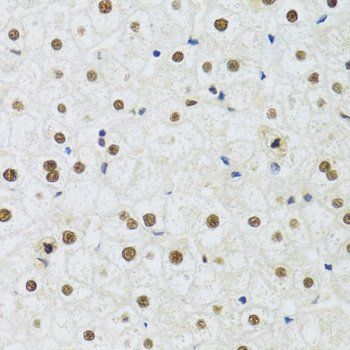
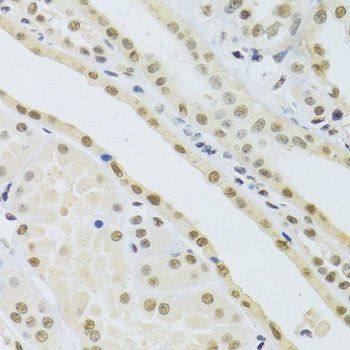
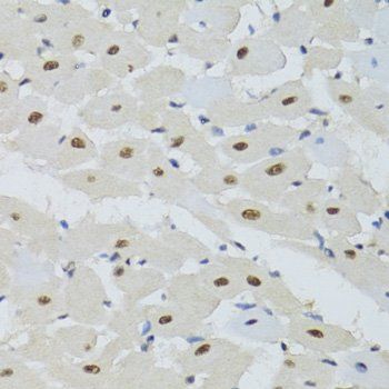
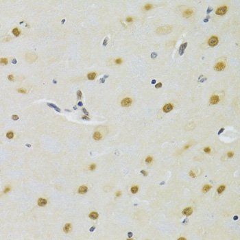
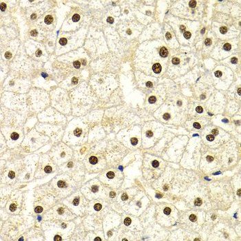
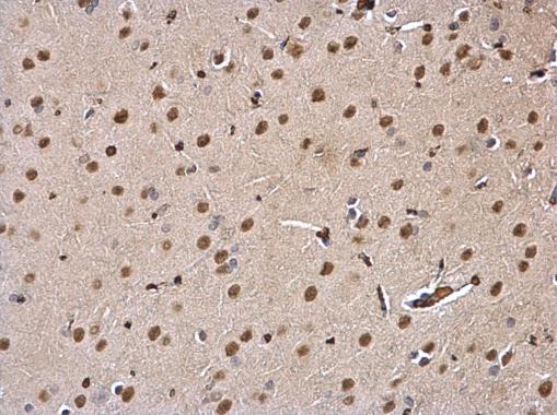
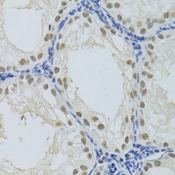
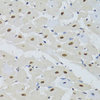
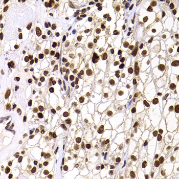
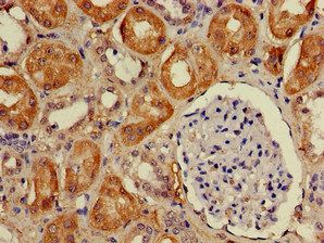
IHC analysis of DDB1 using anti-DDB1 antibody. DDB1 was detected in paraffin-embedded section of Mouse Liver Tissue.
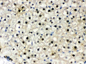
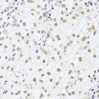
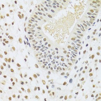
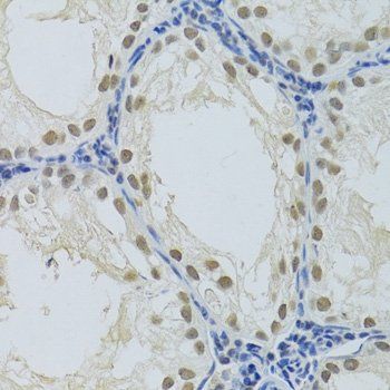
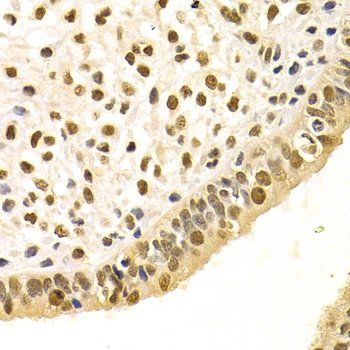
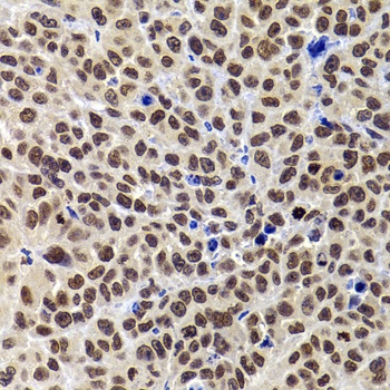
IHC analysis of DDB1 using anti-DDB1 antibody. DDB1 was detected in paraffin-embedded section of Rat Liver Tissue.
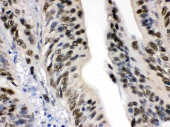
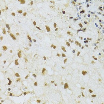
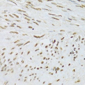
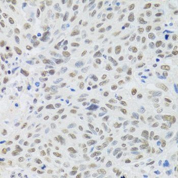
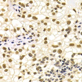
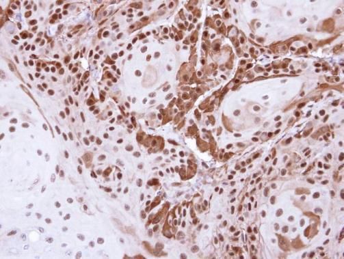
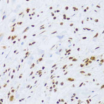
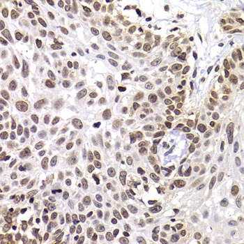
IHC analysis of DDB1 using anti-DDB1 antibody. DDB1 was detected in paraffin-embedded section of Human Intestinal Cancer Tissue.
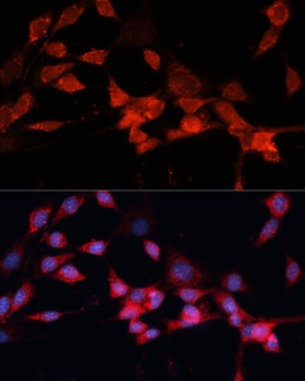
IHC analysis of DDB1 using anti-DDB1 antibody.DDB1 was detected in immunocytochemical section of A549 cell.
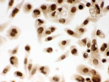
IHC analysis of DDB1 using anti-DDB1 antibody.DDB1 was detected in immunocytochemical section of PC-3 cell.
IHC analysis of DDB1 using anti-DDB1 antibody.DDB1 was detected in immunocytochemical section of SMMC-7721 cell.
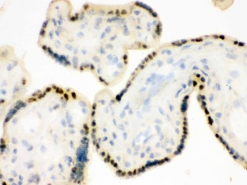
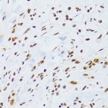
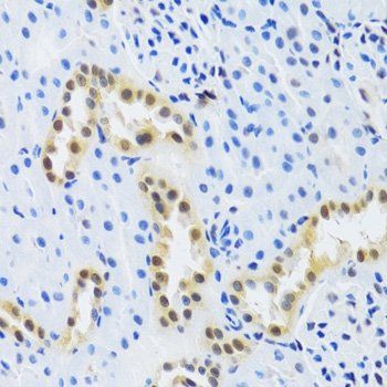
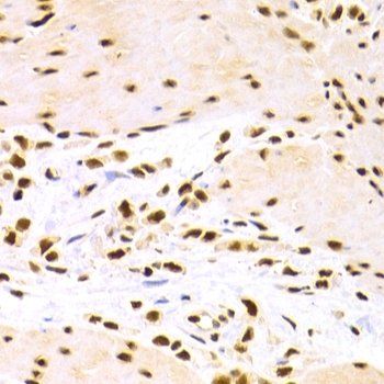
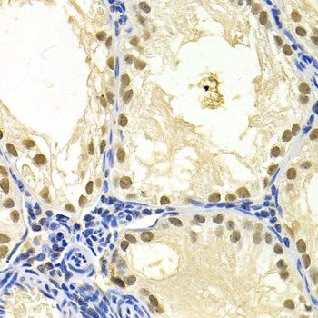
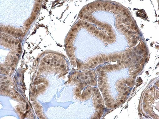
IHC analysis of DDB1 using anti-DDB1 antibody.DDB1 was detected in frozen section of human placenta tissue.
- Item 1 of 18
DDB1 antibody [orb135377]
ICC, IF, IHC, WB
Human, Mouse, Rat
Polyclonal
Unconjugated
50 μl, 100 μl, 200 μl - Item 1 of 11
damage specific DNA binding protein 1 Antibody [orb555881]
ICC, IHC-P, IP, WB
Human, Mouse, Rat
Rabbit
Polyclonal
Unconjugated
100 μl - Item 1 of 6
- Item 1 of 5
- Item 1 of 3
DDA1 antibody [orb52126]
ELISA, IF, IHC, WB
Human, Mouse, Rat
Rabbit
Polyclonal
Unconjugated
100 μg, 50 μg


Submit a review
Filter by Rating

- 5 stars

- 4 stars
- 3 stars
- 2 stars
- 1 stars