You have no items in your shopping cart.
Cart summary
Item 1 of 7
Item 1 of 7
CD9 Antibody
Catalog Number: orb2641126

| Catalog Number | orb2641126 |
|---|---|
| Category | Antibodies |
| Description | CD9 is a type IV transmembrane glycoprotein with four transmembrane domains. CD9 on pre-B cells may play a role in cell-cell adhesion. In addition, CD9 may play a role in signal transduction mediated by interaction with low molecular weight GTP binding proteins. CD9 is expressed on early B cells, eosinophils, basophils and activated T cells and is a major component of the platelet cell surface. It is also expressed on most non-T acute lymphoblastic leukemia cells and on some acute myeloid and chronic lymphoid leukemias. |
| Species/Host | Mouse |
| Clonality | Monoclonal |
| Clone Number | CD9/1619 |
| Tested applications | FACS, IHC-P |
| Reactivity | Human |
| Isotype | Mouse IgG1, kappa |
| Immunogen | Recombinant full-length human protein was used as the immunogen for the CD9 antibody. |
| Antibody Type | Primary Antibody |
| Dilution range | Immunohistochemistry (FFPE): 1-2ug/ml for 30 min at RT,Flow cytometry: 1-2ug/million cells |
| Purity | Protein G affinity chromatography |
| Conjugation | Unconjugated |
| Formula | 0.2 mg/ml in 1X PBS with 0.1 mg/ml BSA (US sourced) and 0.05% sodium azide |
| Hazard Information | This CD9 antibody is available for research use only. |
| UniProt ID | P21926 |
| Storage | Maintain refrigerated at 2-8°C for up to 2 weeks. For long term storage store at -20°C in small aliquots to prevent freeze-thaw cycles. |
| Buffer/Preservatives | 0.2 mg/ml in 1X PBS with 0.1 mg/ml rAlbumin (US sourced) and 0.05% sodium azide |
| Note | For research use only |
| Application notes | Titering of the CD9 antibody may be required for optimal performance. |
| Expiration Date | 12 months from date of receipt. |

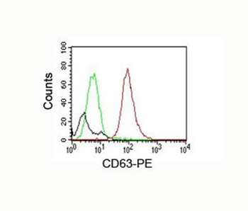
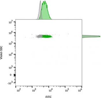
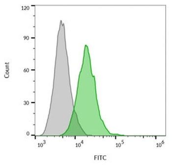
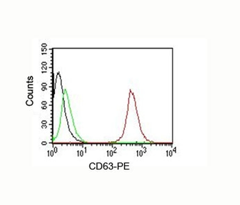
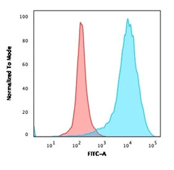
Flow cytometry staining of human MCF7 cells with CD9 antibody (clone CD9/1619); Gray = unstained, Red = CD9 antibody.
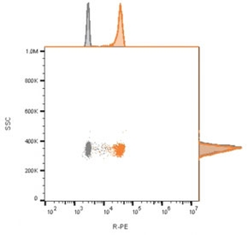
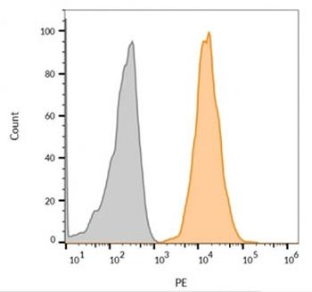
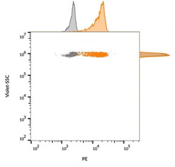
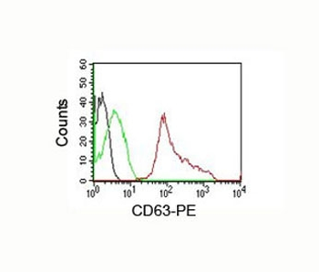
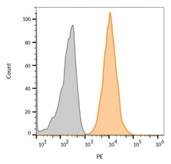
FACS staining of bead-bound exosomes derived from human MCF7 cells: Gray = unstained, Orange = CF568 labeled CD63 antibody (clone CD9/1619).

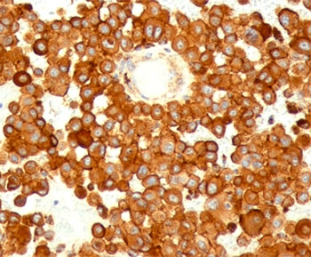
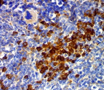
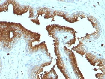
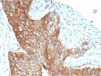
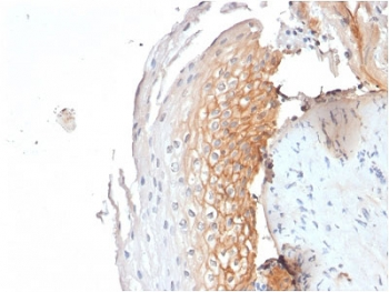
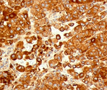
IHC staining of FFPE human basal cell carcinoma with CD9 antibody (clone CD9/1619). HIER: boil tissue sections in pH6, 10mM citrate buffer, for 10-20 min followed by cooling at RT for 20 min.

IHC staining of FFPE human cervical carcinoma with CD9 antibody (clone CD9/1619). HIER: boil tissue sections in pH6, 10mM citrate buffer, for 10-20 min followed by cooling at RT for 20 min.
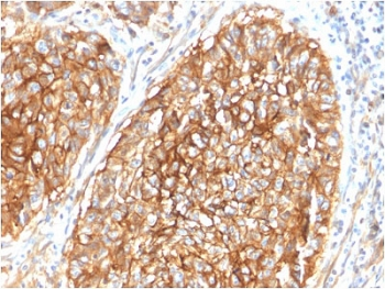
IHC staining of FFPE human lung carcinoma with CD9 antibody (clone CD9/1619). HIER: boil tissue sections in pH6, 10mM citrate buffer, for 10-20 min followed by cooling at RT for 20 min.

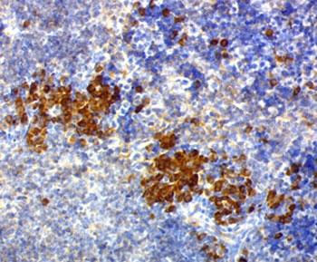
IHC staining of FFPE human tonsil with CD9 antibody (clone CD9/1619). HIER: boil tissue sections in pH6, 10mM citrate buffer, for 10-20 min followed by cooling at RT for 20 min.
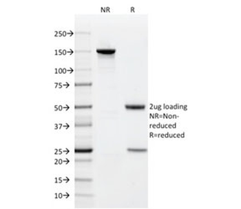
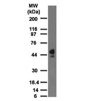
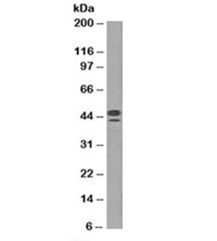
SDS-PAGE analysis of purified, BSA-free CD9 antibody (clone CD9/1619) as confirmation of integrity and purity.
- Item 1 of 11
- Item 1 of 11
CD63 Antibody / LAMP-3 [orb2637613]
FACS, IF, IHC-P, WB
Human, Mouse
Mouse
Monoclonal
Unconjugated
100 μg - Item 1 of 7
- Item 1 of 7
- Item 1 of 7