You have no items in your shopping cart.
Cart summary
Item 1 of 11
Item 1 of 11
CD63 Antibody / LAMP-3
Catalog Number: orb2637613

| Catalog Number | orb2637613 |
|---|---|
| Category | Antibodies |
| Description | The tetraspanins are integral membrane proteins expressed on cell surface and granular membranes of hematopoietic cells and are components of multi-molecular complexes with specific integrins. The tetraspanin CD63 is a lysosomal membrane glycoprotein that translocates to the plasma membrane after platelet activation. CD63 is expressed on activated platelets, monocytes and macrophages, and is weakly expressed on granulocytes, T cell and B cells. It is located on the basophilic granule membranes and on the plasma membranes of lymphocytes and granulocytes. CD63 is a member of the TM4 superfamily of leukocyte glycoproteins that includes CD9, CD37 and CD53, which contain four transmembrane regions. CD63 may play a role in phagocytic and intracellular lysosome-phagosome fusion events. CD63 deficiency is associated with Hermansky-Pudlak syndrome and is strongly expressed during the early stages of melanoma progression. |
| Clonality | Monoclonal |
| Species/Host | Mouse |
| Isotype | Mouse IgG1, kappa |
| Conjugation | Unconjugated |
| Reactivity | Human, Mouse |
| Buffer/Preservatives | 0.2 mg/ml in 1X PBS with 0.1 mg/ml rAlbumin (US sourced) and 0.05% sodium azide |
| Purity | Protein G affinity chromatography |
| Immunogen | Full length human CD63 was used as the immunogen for this antibody. |
| Tested applications | FACS, IF, IHC-P, WB |
| Dilution range | Flow cytometry: 1-2ug/million cells,Immunofluorescence: 1-2ug/ml,Western blot: 1-2ug/ml,Immunohistochemistry (FFPE): 1-2ug/ml for 30 min at RT |
| Application notes | The concentration stated for each application is a general starting point. Variations in protocols, secondaries and substrates may require the antibody to be titered up or down for optimal performance.1. Staining of formalin-fixed tissues is enhanced by boiling tissue sections in pH 9 10mM Tris with 1mM EDTA for 10-20 min followed by cooling at RT for 20 minutes.2. The prediluted format is supplied in a dropper bottle and is optimized for use in IHC. After epitope retrieval step (if required), drip mAb solution onto the tissue section and incubate at RT for 30 min. |
| Antibody Type | Primary Antibody |
| Clone Number | MX-49.129.5 |
| Formula | 0.2 mg/ml in 1X PBS with 0.1 mg/ml BSA (US sourced) and 0.05% sodium azide |
| Storage | Maintain refrigerated at 2-8°C for up to 2 weeks. For long term storage store at -20°C in small aliquots to prevent freeze-thaw cycles. |
| Hazard Information | This CD63 antibody is available for research use only. |
| Note | For research use only |
| Entrez | 967 |
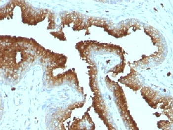
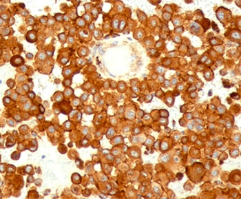
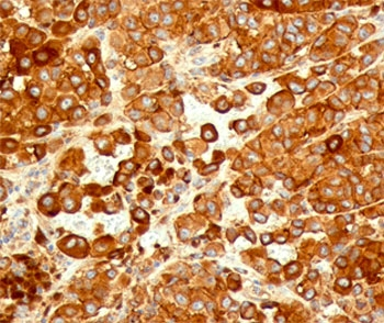
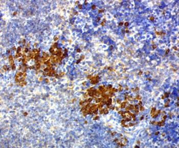
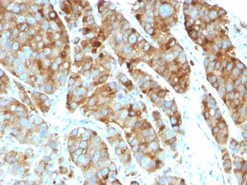
IHC testing of FFPE prostate carcinoma with CD63 antibody (clone MX-49.129.5).

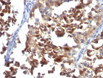
IHC testing of FFPE human melanoma stained with CD63 antibody (clone MX49.129.5).

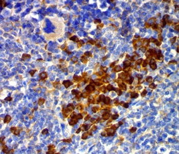
IHC testing of FFPE mouse spleen stained with CD63 antibody (clone MX49.129.5).
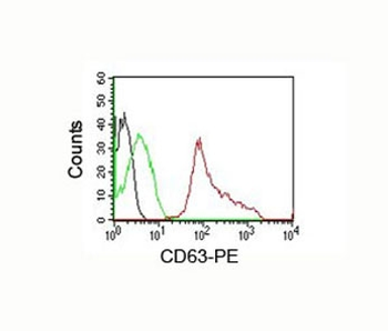
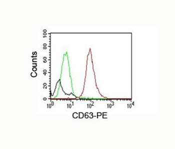
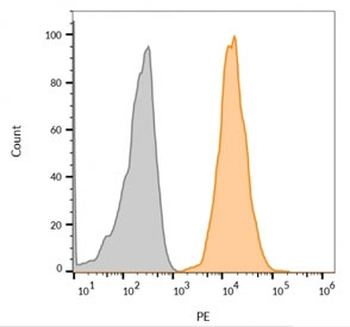
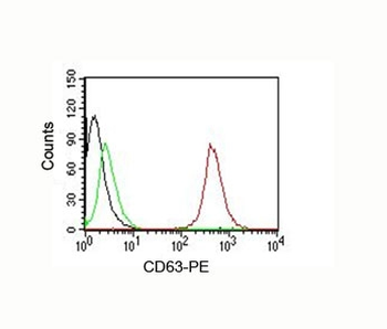
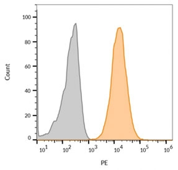
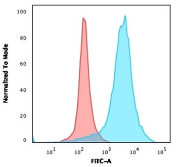
FACS testing of human PBMC: Black = cells alone; Green = isotype control; Red = CD63 antibody PE conjugate

FACS testing of mouse NIH3T3: Black = cells alone; Green = isotype control; Red = CD63 antibody PE conjugate

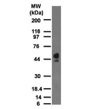
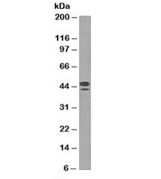
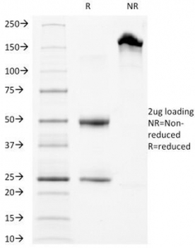
Western blot testing of human spleen lysate with CD63 antibody at 2 ug/ml (clone MX-49.129.5).
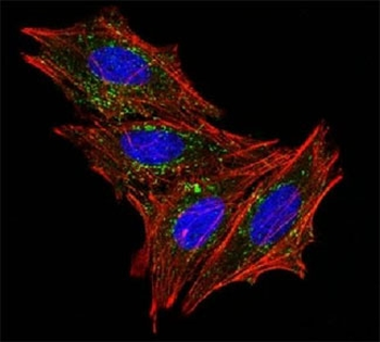
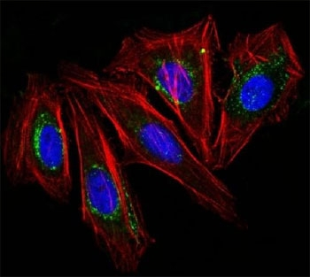
Immunofluorescence testing of HeLa cells with Alexa Fluor 488 conjugated CD63 antibody (green). F-actin filaments are labeled with Dylight 554 phalloidin (red); nuclei stained with DAPI (blue).
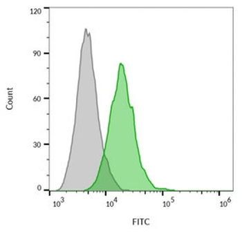
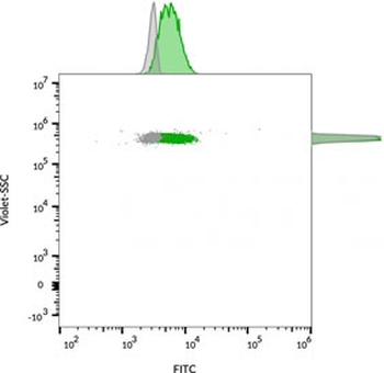
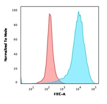
FACS staining of human MCF7 cells: Gray = unstained, Green = CF488 labeled CD63 antibody.

FACS staining of bead-bound exosomes derived from human MCF7 cells: Gray = unstained, Green = CF488 labeled CD63 antibody.

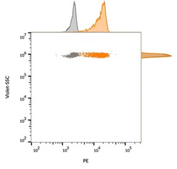
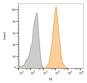
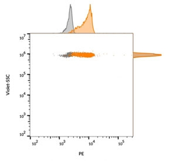
FACS staining of human MCF7 cells: Gray = unstained, Orange = CF568 labeled CD63 antibody.

FACS staining of bead-bound exosomes derived from human MCF7 cells: Gray = unstained, Orange = CF568 labeled CD63 antibody.
- Item 1 of 11
- Item 1 of 7
- Item 1 of 7
- Item 1 of 6
- Item 1 of 6