You have no items in your shopping cart.
Cart summary
Item 1 of 5
Item 1 of 5
CD79a Antibody
Catalog Number: orb2637630

| Catalog Number | orb2637630 |
|---|---|
| Category | Antibodies |
| Description | CD79a is required in cooperation with CD79b for initiation of the signal transduction cascade activated by binding of antigen to the B-cell antigen receptor complex (BCR) which leads to internalization of the complex, trafficking to late endosomes and antigen presentation. Also required for BCR surface expression and for efficient differentiation of pro- and pre-B-cells. Stimulates SYK autophosphorylation and activation. Binds to BLNK, bringing BLNK into proximity with SYK and allowing SYK to phosphorylate BLNK. Also interacts with and increases activity of some Src-family tyrosine kinases. Represses BCR signaling during development of immature B-cells. [UniProt] |
| Species/Host | Mouse |
| Clonality | Monoclonal |
| Clone Number | HM47/A9 |
| Tested applications | FACS, IF, IHC-P, WB |
| Reactivity | Human, Mouse, Rat |
| Isotype | Mouse IgG1, kappa |
| Immunogen | A synthetic peptide corresponding to aa 202-216 (GTYQDVGSLNIADVQ) of human CD79a protein was used as the immunogen for this antibody. |
| Antibody Type | Primary Antibody |
| Dilution range | Flow cytometry: 1-2ug/million cells,Immunofluorescence: 1-2ug/ml,Western blot: 1-2ug/ml,Immunohistochemistry (FFPE): 1-2ug/ml for 30 min at RT |
| Purity | Protein G affinity chromatography |
| Conjugation | Unconjugated |
| Formula | 0.2 mg/ml in 1X PBS with 0.1 mg/ml BSA (US sourced) and 0.05% sodium azide |
| Hazard Information | This CD79a antibody is available for research use only. |
| UniProt ID | P11912 |
| Storage | Maintain refrigerated at 2-8°C for up to 2 weeks. For long term storage store at -20°C in small aliquots to prevent freeze-thaw cycles. |
| Buffer/Preservatives | 0.2 mg/ml in 1X PBS with 0.1 mg/ml rAlbumin (US sourced) and 0.05% sodium azide |
| Note | For research use only |
| Application notes | The concentration stated for each application is a general starting point. Variations in protocols, secondaries and substrates may require the antibody to be titered up or down for optimal performance.1. Staining of FFPE tissues requires boiling sections in pH 9 10mM Tris with 1mM EDTA for 10-20 min followed by cooling at RT for 20 min.2. The prediluted format is supplied in a dropper bottle and is optimized for use in IHC. After epitope retrieval step (if required), drip mAb solution onto the tissue section and incubate at RT for 30 min. |
| Expiration Date | 12 months from date of receipt. |
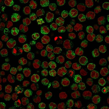
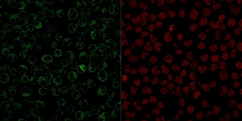
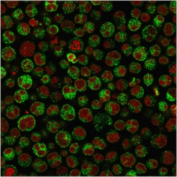
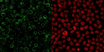
Immunofluorescent staining of PFA-fixed human Raji cells with CD79a antibody (clone HM47/A9, green) and Reddot nuclear stain (red).

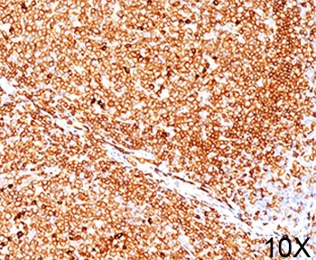
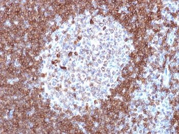
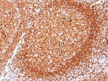
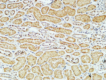
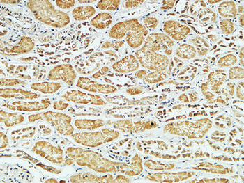
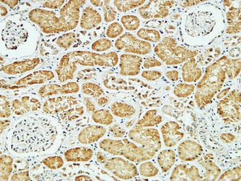
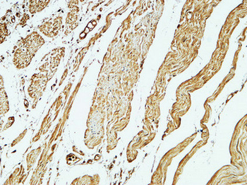
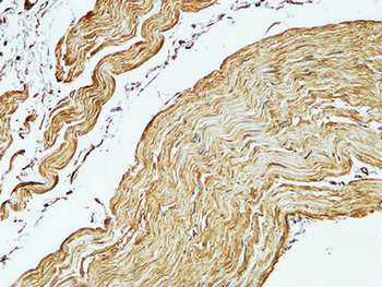
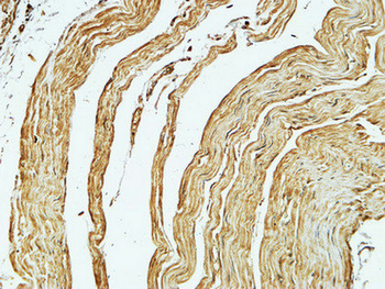
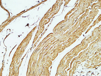
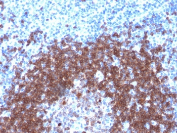
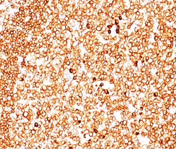
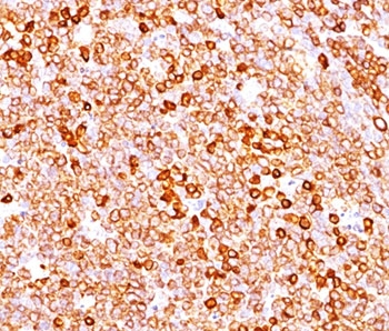
IHC testing of FFPE human tonsil (10X) stained with CD79a antibody (clone HM47/A9).
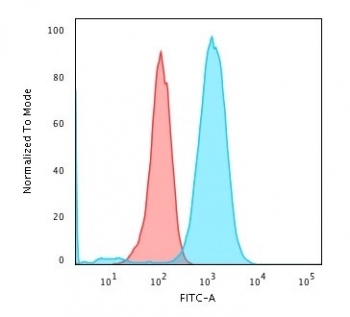
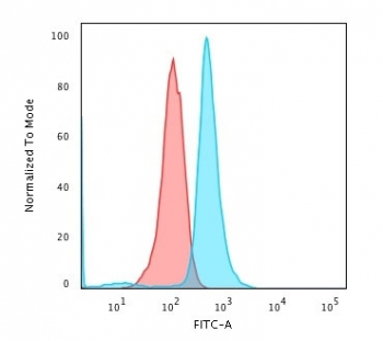
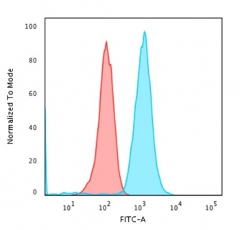
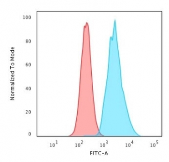
Flow cytometry testing of human Raji cells with CD79a antibody (clone HM47/A9); Red = isotype control, Blue = CD79a antibody.

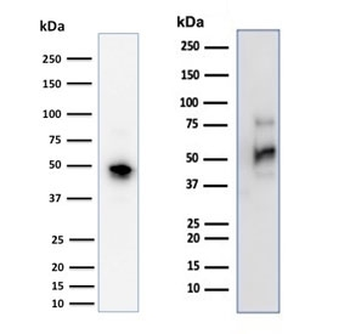
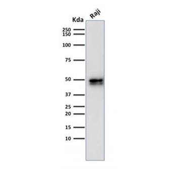
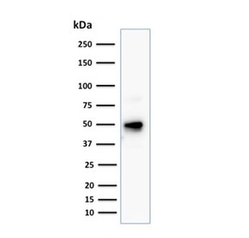
Western blot testing of human Raji cell lysate with CD79a antibody (clone HM47/A9). Expected molecular weight: 25-47 kDa depending on glycosylation level.
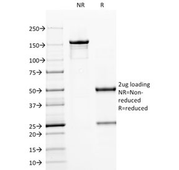
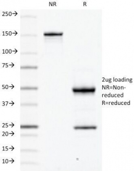
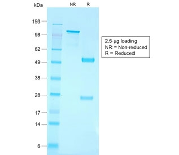
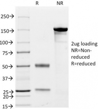
SDS-PAGE analysis of purified, BSA-free CD79a antibody (clone HM47/A9) as confirmation of integrity and purity.
- Item 1 of 7
- Item 1 of 7
- Item 1 of 7
- Item 1 of 6
- Item 1 of 6
CD79a Antibody Cocktail [orb248285]
FACS, IF, IHC-P, WB
Human, Mouse, Rat
Mouse
Monoclonal
Unconjugated
20 μg