You have no items in your shopping cart.
Cart summary
Item 1 of 6
Item 1 of 6
CD63 Antibody
Catalog Number: orb1271031

| Catalog Number | orb1271031 |
|---|---|
| Category | Antibodies |
| Description | CD63 Antibody |
| Target | CD63 |
| Clonality | Polyclonal |
| Isotype | Rabbit Ig |
| Conjugation | Unconjugated |
| Reactivity | Human |
| Form/Appearance | Liquid |
| Concentration | batch dependent |
| Buffer/Preservatives | Supplied in PBS with 0.09% (W/V) sodium azide. |
| Purification | This antibody is purified through a protein A column, followed by peptide affinity purification. |
| Immunogen | This CD63 antibody is generated from rabbits immunized with a KLH conjugated synthetic peptide between 163-190 amino acids from the C-terminal region of human CD63. |
| UniProt ID | P08962 |
| MW | 26 kDa |
| Tested applications | FC, IHC-P, WB |
| Application notes | For IHC-P starting dilution is: 1:25For FACS starting dilution is: 1:25For WB starting dilution is: 1:1000 |
| Antibody Type | Primary Antibody |
| Storage | Maintain refrigerated at 2-8°C for up to 2 weeks. For long term storage store at -20°C in small aliquots to prevent freeze-thaw cycles. |
| Alternative names | CD63 antigen, Granulophysin, Lysosomal-associated Read more... |
| Note | For research use only |
| NCBI | P08962 |
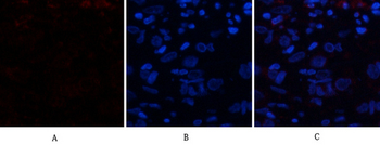
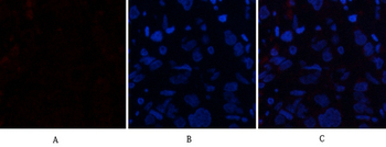
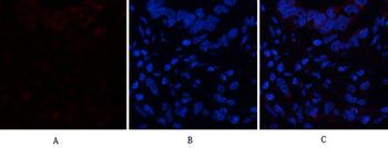
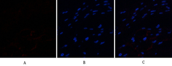
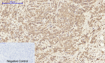
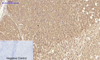
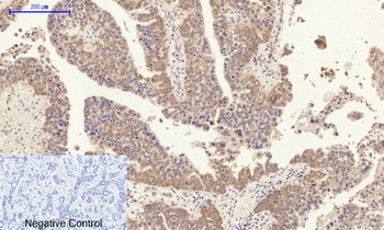
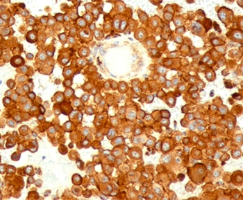
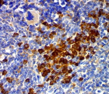
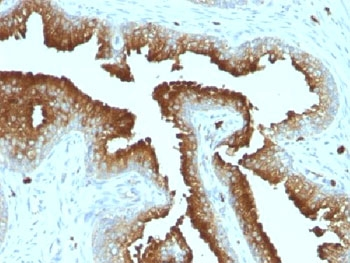
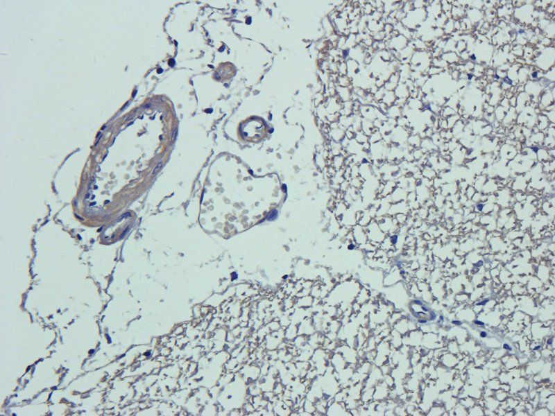
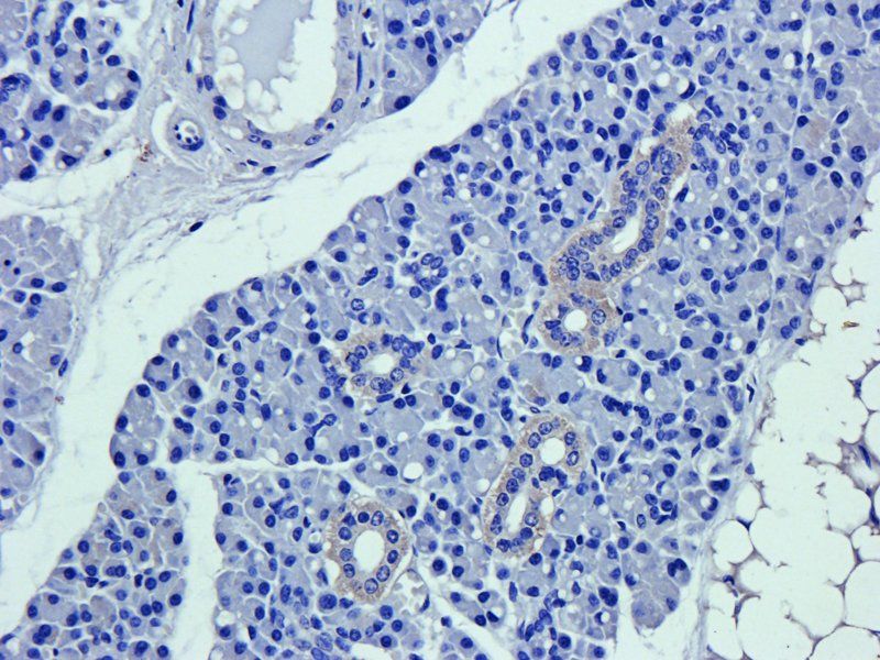
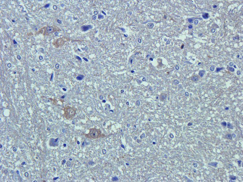
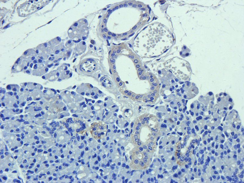
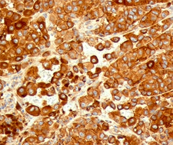
Antibody staining CD63 in human melanoma tissue sections by Immunohistochemistry (IHC-P - paraformaldehyde-fixed, paraffin-embedded sections).
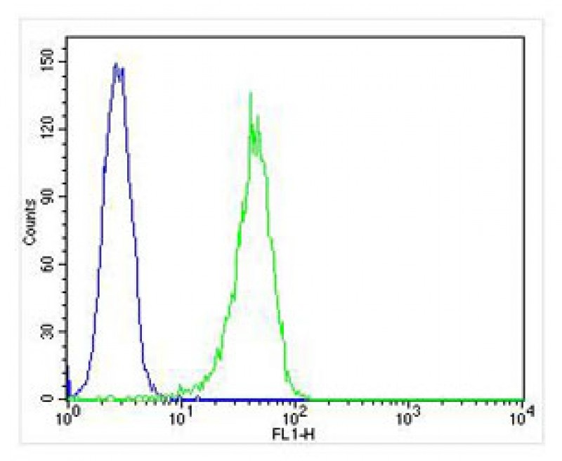
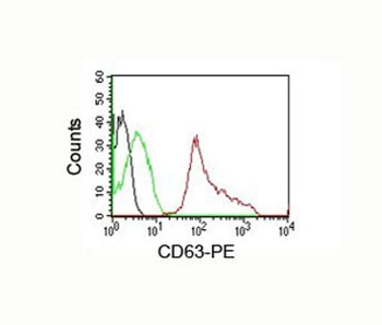
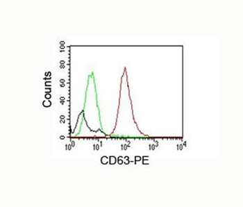
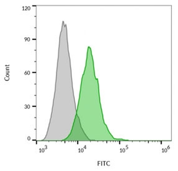
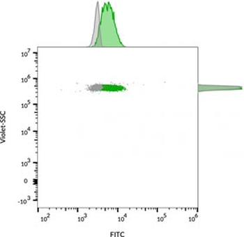
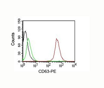
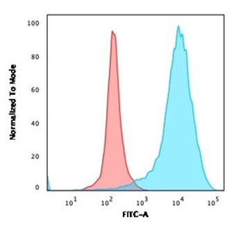
Overlay histogram showing HL-60 cells stained with Antibody (green line). The cells were fixed with 4% paraformaldehyde (10 min) and then permeabilized with 90% methanol for 10 min. The cells were then icubated in 2% bovine serum albumin to block non-specific protein-protein interactions followed by the antibody (1:25 dilution) for 60 min at 37°C. The secondary antibody used was Alexa Fluor 488 goat anti-rabbit lgG (H + L) at 1/400 dilution for 40 min at 37°C. Isotype control antibody (blue line) was rabbit IgG1 (1ug/1x10^6 cells) used under the same conditions. Acquisition of > 10000 events was performed.
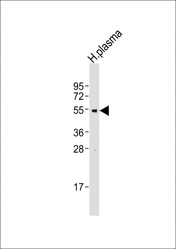
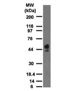
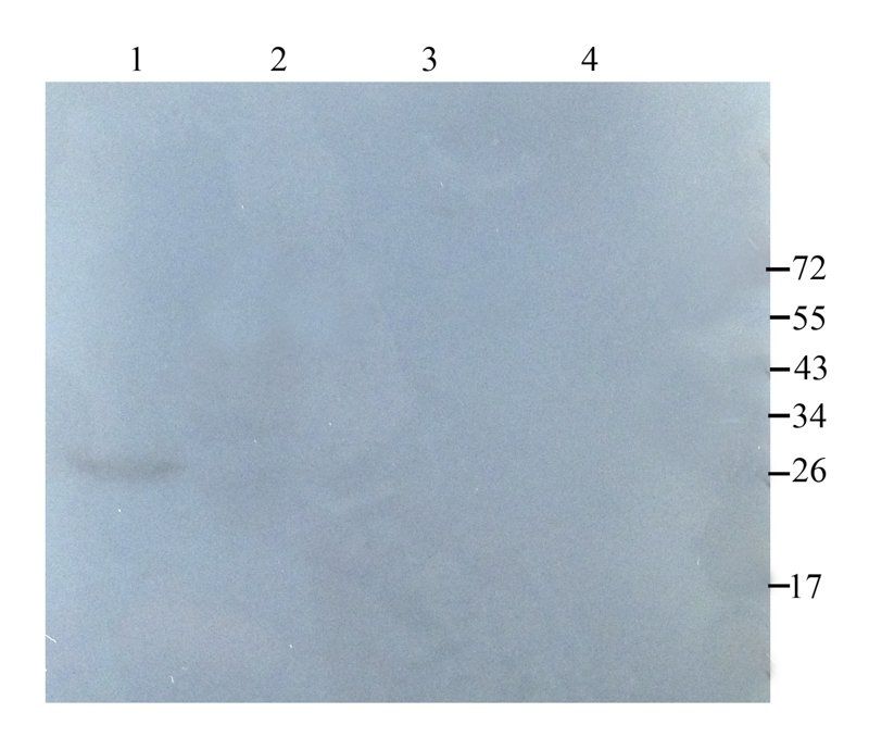
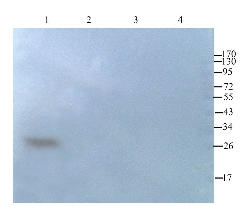
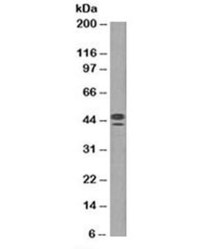
Western Blot at 1:2000 dilution + human plasma lysates Lysates/proteins at 20 ug per lane.
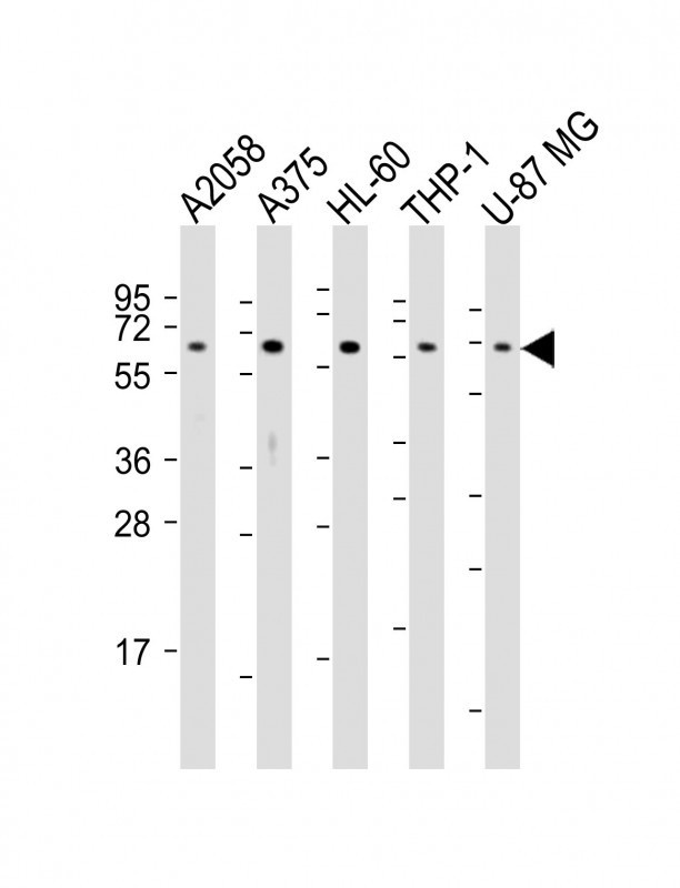
Western Blot at 1:2000 dilution Lane 1: A2058 whole cell lysates Lane 2: A375 whole cell lysates Lane 3: HL-60 whole cell lysates Lane 4: THP-1 whole cell lysates Lane 5: U-87 MG whole cell lysates Lysates/proteins at 20 ug per lane.
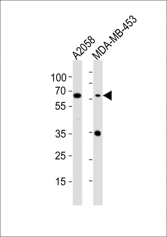
Western Blot at 1:1000 dilution Lane 1: A2058 whole cell lysates Lane 2: MDA-MB-453 whole cell lysates Lysates/proteins at 20 ug per lane.
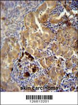
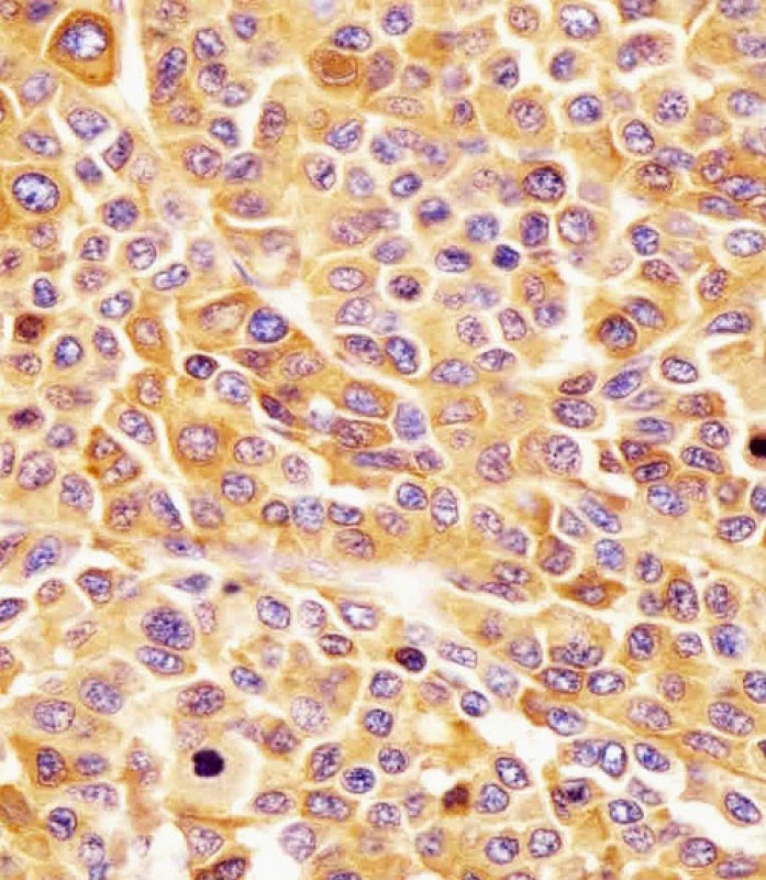
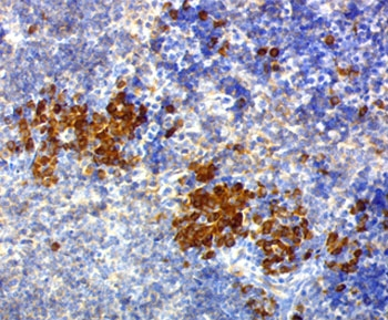
CD63 Antibody immunohistochemistry analysis in formalin fixed and paraffin embedded human skin carcinoma followed by peroxidase conjugation of the secondary antibody and DAB staining.
- Item 1 of 11
CD63 Antibody / LAMP-3 [orb749338]
FACS, IF, IHC-P, WB
Human, Mouse
Mouse
Monoclonal
Unconjugated
20 μg, 100 μg - Item 1 of 6
- Item 1 of 7
CD63 Antibody / LAMP-3 [orb749335]
FACS, IF, IHC-P
Human, Mouse
Mouse
Monoclonal
Unconjugated
20 μg, 100 μg - Item 1 of 7
- Item 1 of 7
![CD63 Antibody [MX-49.129.5]](/images//pub/media/catalog/product/NewWebsite/15/orb1252797_1.jpg)
![CD63 Antibody [MX-49.129.5]](/images/pub/media/catalog/product/NewWebsite/15/orb1252797_2.jpg)
![CD63 Antibody [MX-49.129.5]](/images/pub/media/catalog/product/NewWebsite/15/orb1252797_3.jpg)
![CD63 Antibody [MX-49.129.5]](/images/pub/media/catalog/product/NewWebsite/15/orb1252797_4.jpg)
![CD63 Antibody [MX-49.129.5]](/images/pub/media/catalog/product/NewWebsite/15/orb1252797_5.jpg)
![CD63 Antibody [MX-49.129.5]](/images/pub/media/catalog/product/NewWebsite/15/orb1252797_6.jpg)
![CD63 Antibody [MX-49.129.5]](/images/pub/media/catalog/product/NewWebsite/15/orb1252797_7.jpg)