You have no items in your shopping cart.
Cart summary
Item 1 of 1
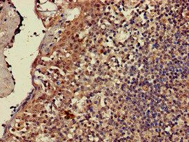
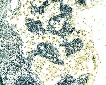
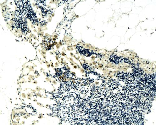
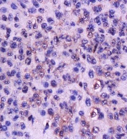
CD3 antibody
Catalog Number: orb44679
| Catalog Number | orb44679 |
|---|---|
| Category | Antibodies |
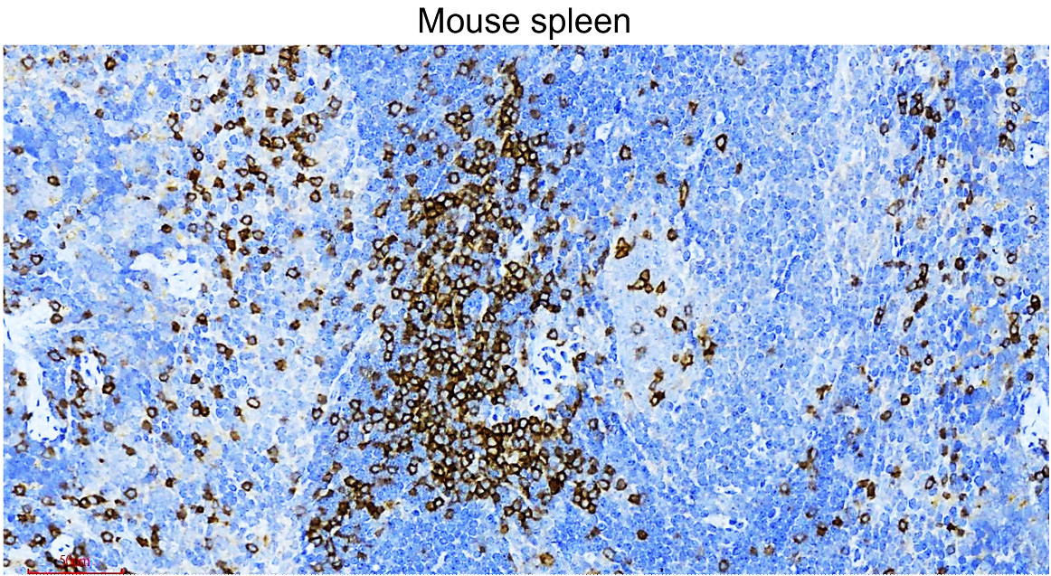
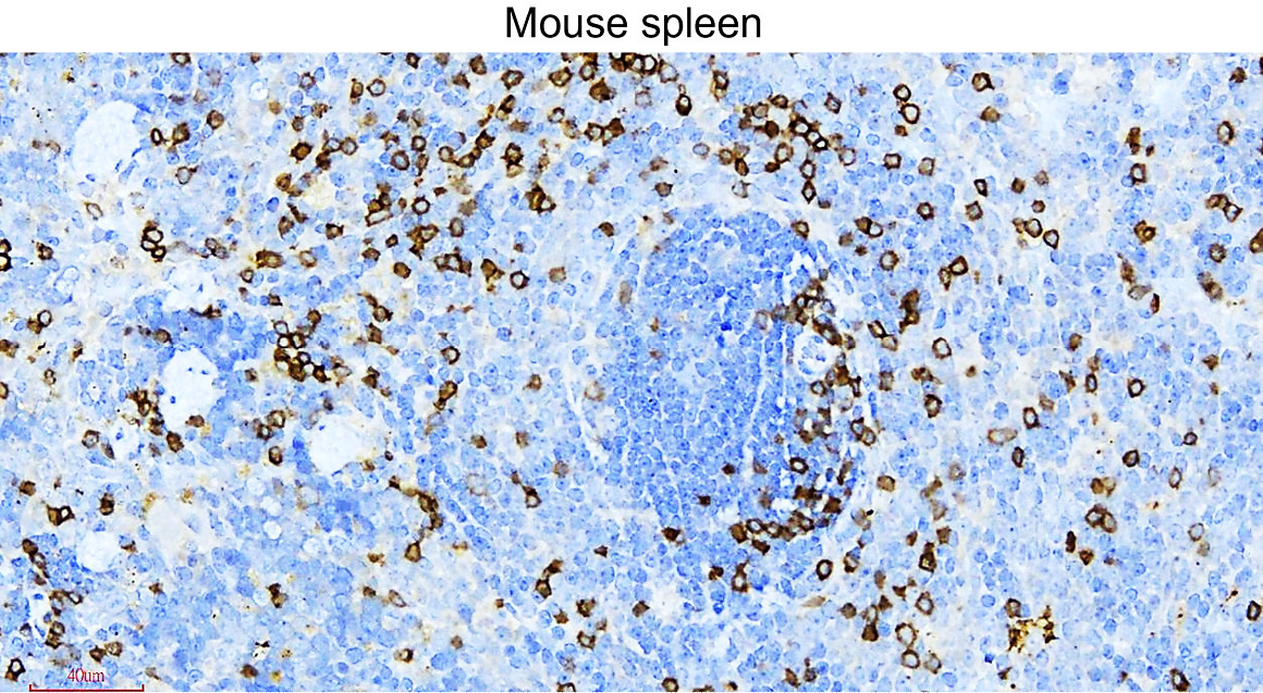
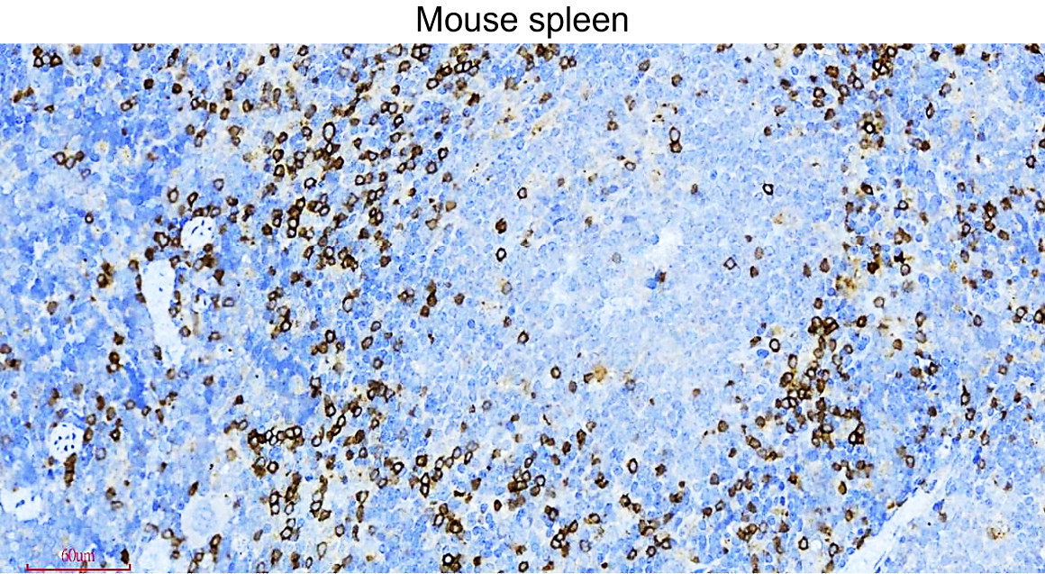
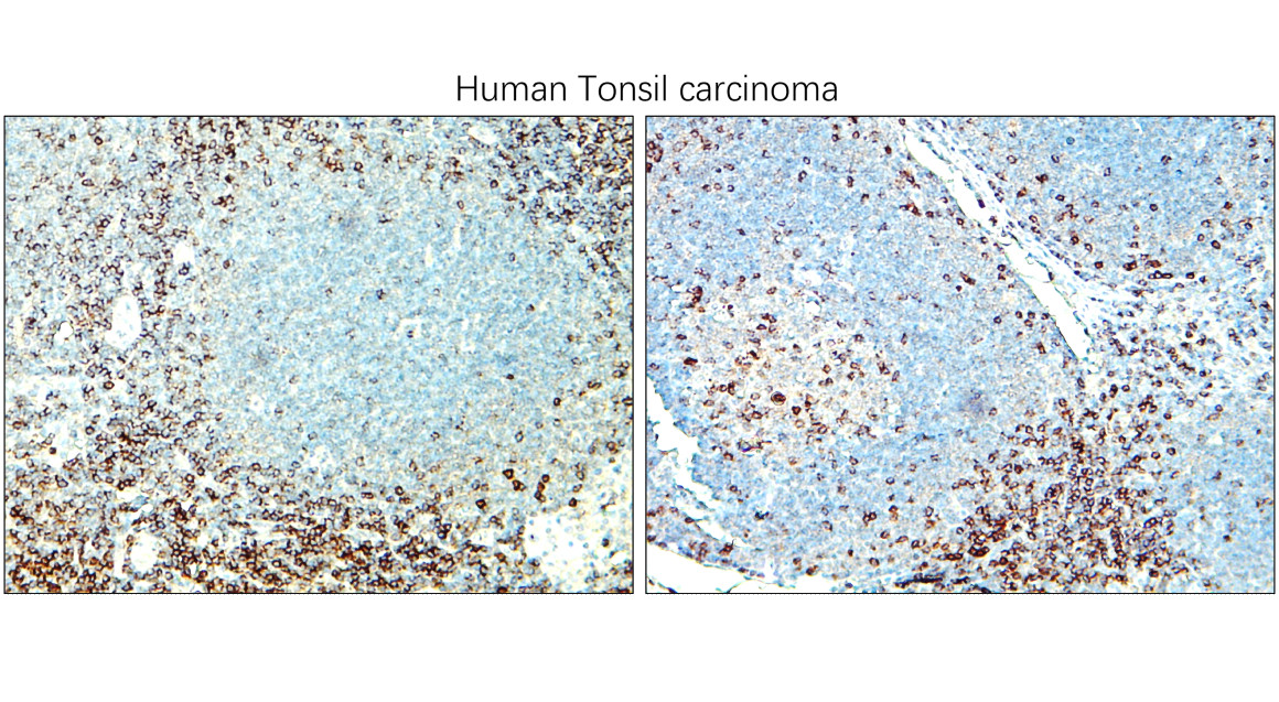
| Description | Mouse monoclonal antibody to CD3 |
| Clonality | Monoclonal |
| Clone Number | APA1/1 |
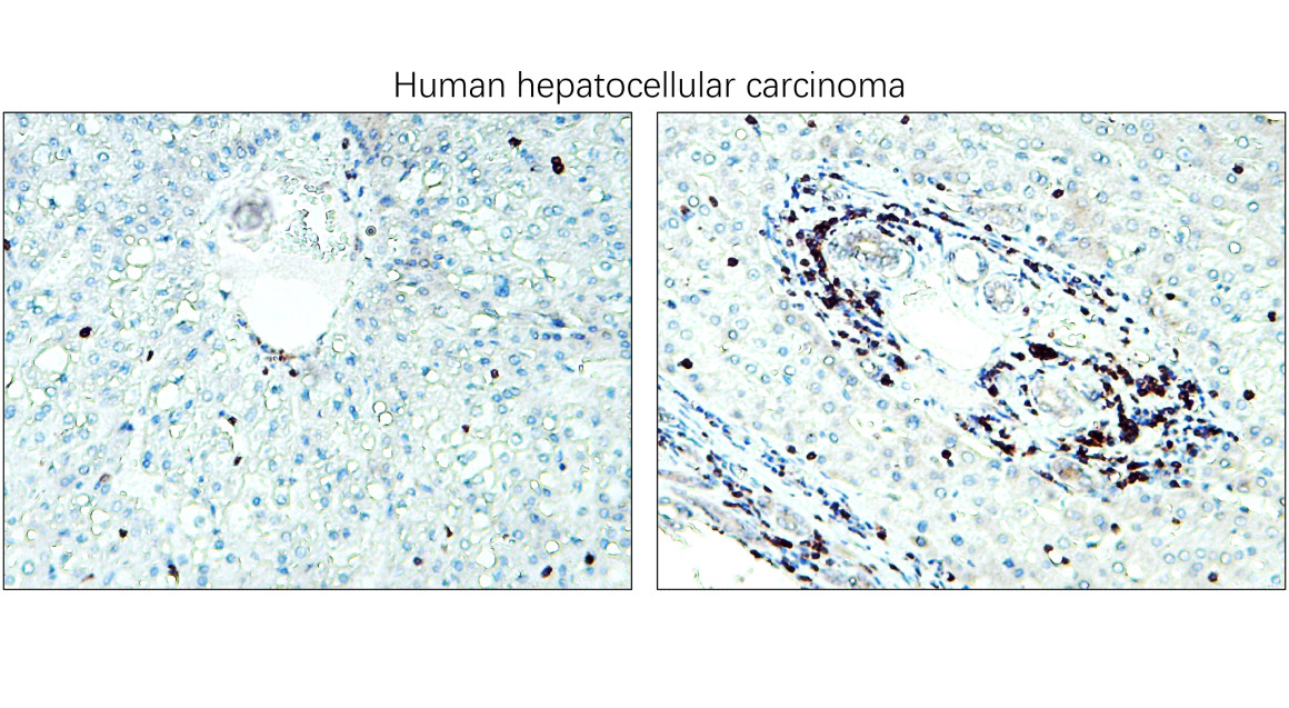
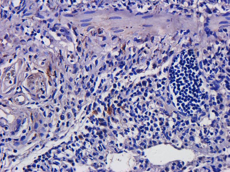
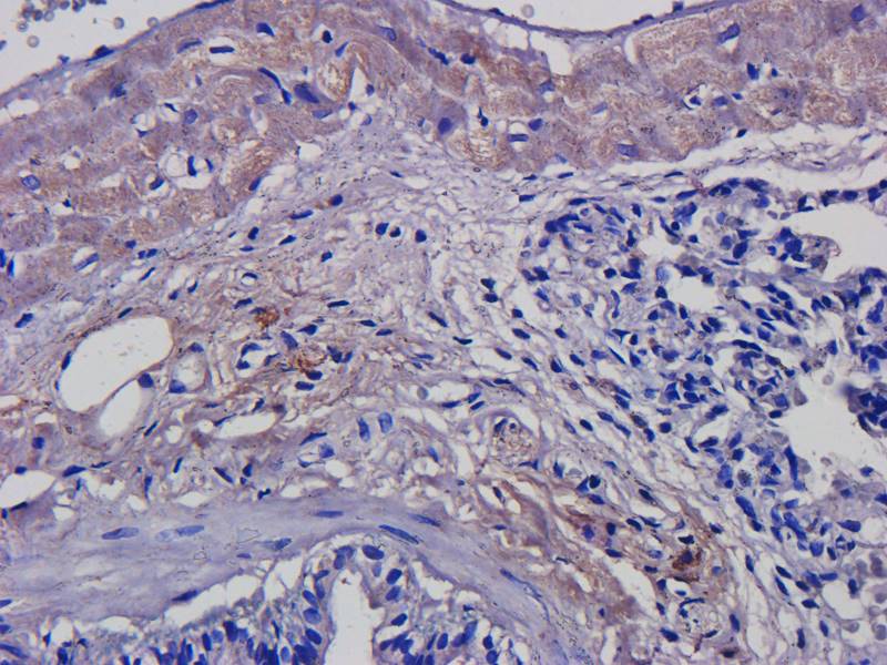
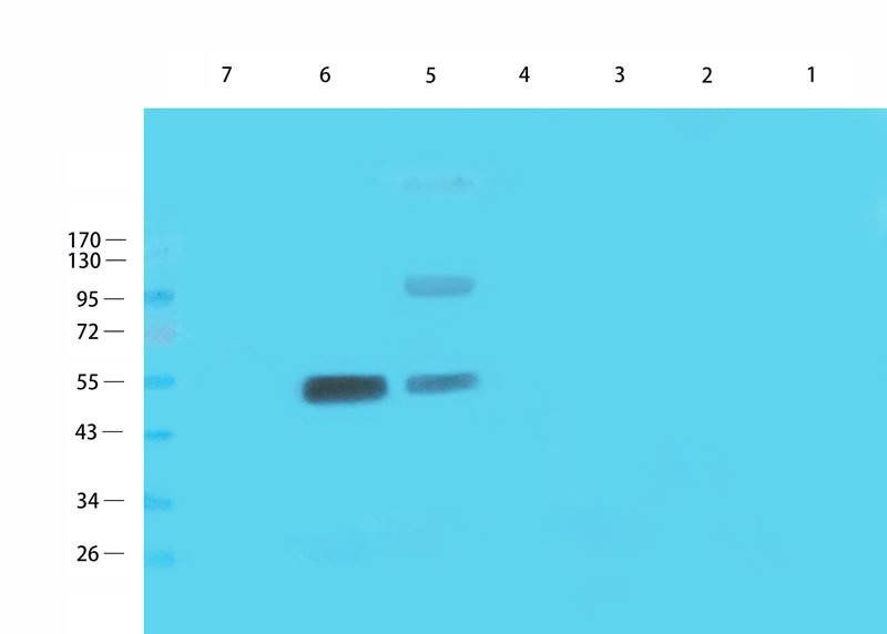
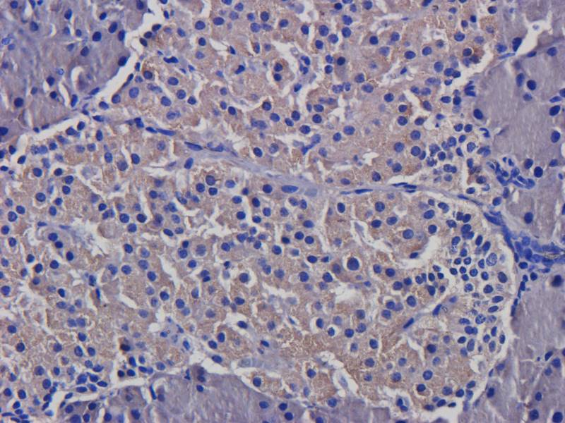
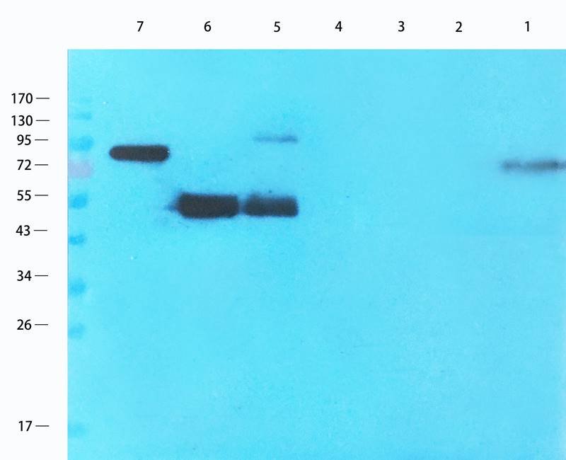
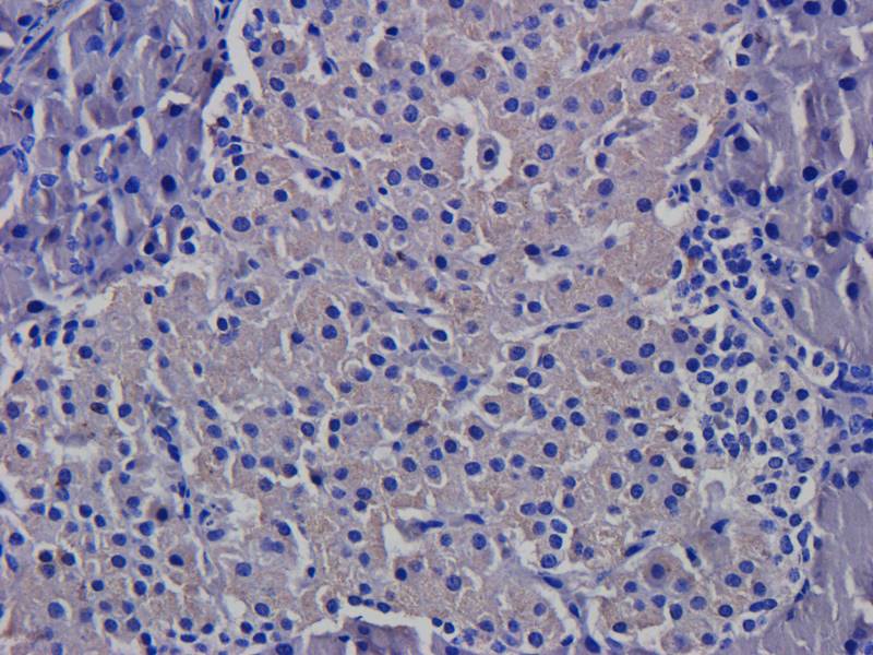
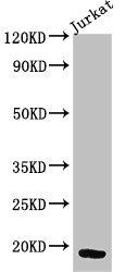
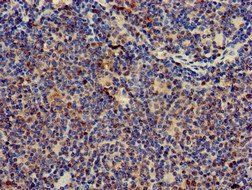
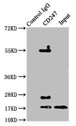
| Tested applications | FC, ICC, IHC-Fr, IP, WB |
| Reactivity | Human, Mouse |
| Isotype | Mouse IgG1 |
| Immunogen | Purified human CD3 proteins isolated from thymus |
| Concentration | 1 mg/ml |
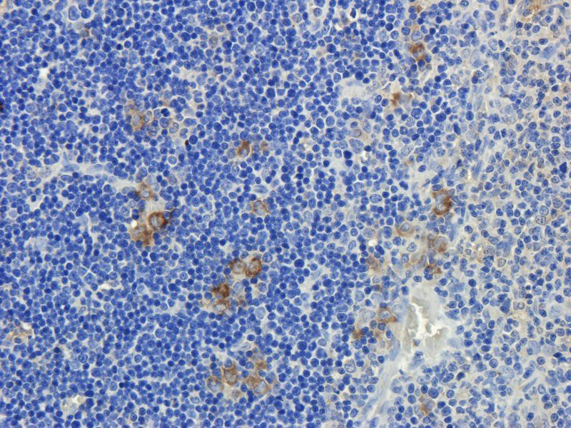
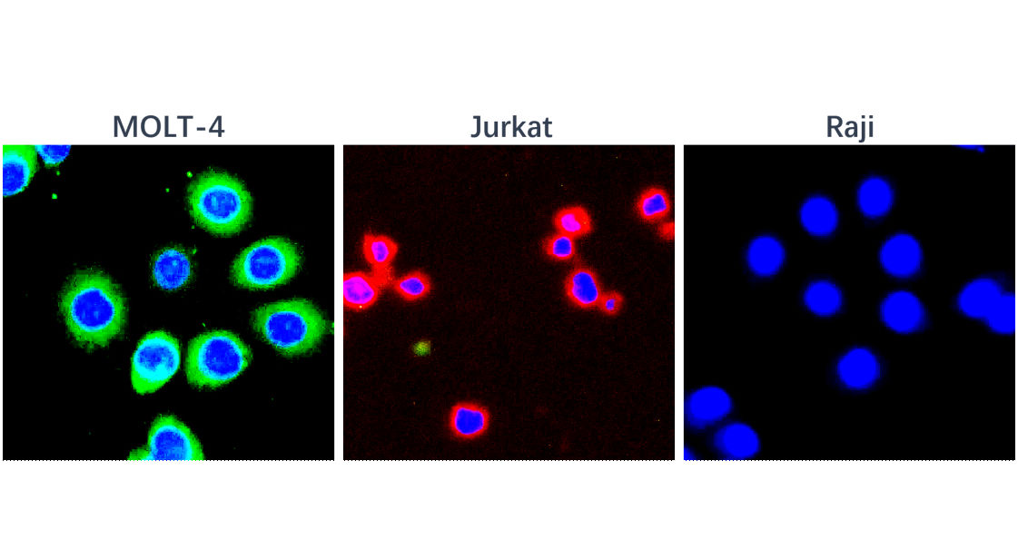
| Dilution range | Flow cytometry: Intracellular staining; recommended dilution: 1-4 µg/ml; positive control: human T cells stimulated with anti-CD3 (MEM-57) antibody (1 μg/ml). Sample preparation: At the end of stimulation of T cells, perform staining of surface markers (if required) in PBS + 0.1% BSA for 20 min. on ice. Wash with PBS and fix with 2% formaldehyde, 30 min on ice. Wash with PBS and incubate in PBS + 0,1% saponine, 5 min. RT. Incubate the cells in PBS + 1% BSA + 0.03% saponine, 15 min. on ice. Incubate with fluorescence-labeled APA1/1 antibody (1-2 μg/ml) in PBS + 1% BSA + 0.03% saponine in dark, 20 min. RT. Wash with PBS + 1% BSA + 0.03% saponine, resuspend in PBS. Immunocytochemistry: Fixed and permeabilised cells. The antibody can distinguish TCR-stimulated from non-stimulated cells. |
| Purity | Purified by protein-A affinity chromatography. |
| Conjugation | Unconjugated |
| Target | CD3 epsilon (activation epitope) |
| Entrez | 916 |
| UniProt ID | P07766 |
| Storage | Store at 2-8°C. Do not freeze. |
| Buffer/Preservatives | Phosphate buffered saline (PBS), pH 7.4, 15 mM sodium azide |
| Alternative names | anti CD 3 antibody, anti Cd247 antibody, anti CD24 Read more... |
| Note | For research use only |
| Application notes | Flow cytometry: Intracellular staining; recommended dilution: 1-4 µg/ml; positive control: human T cells stimulated with anti-CD3 (MEM-57) antibody (1 μg/ml). Sample preparation: At the end of stimulation of T cells, perform staining of surface markers (if required) in PBS + 0.1% BSA for 20 min. on ice. Wash with PBS and fix with 2% formaldehyde, 30 min on ice. Wash with PBS and incubate in PBS + 0,1% saponine, 5 min. RT. Incubate the cells in PBS + 1% BSA + 0.03% saponine, 15 min. on ice. Incubate with fluorescence-labeled APA1/1 antibody (1-2 μg/ml) in PBS + 1% BSA + 0.03% saponine in dark, 20 min. RT. Wash with PBS + 1% BSA + 0.03% saponine, resuspend in PBS. Immunocytochemistry: Fixed and permeabilised cells. The antibody can distinguish TCR-stimulated from non-stimulated cells. |
| Expiration Date | 12 months from date of receipt. |
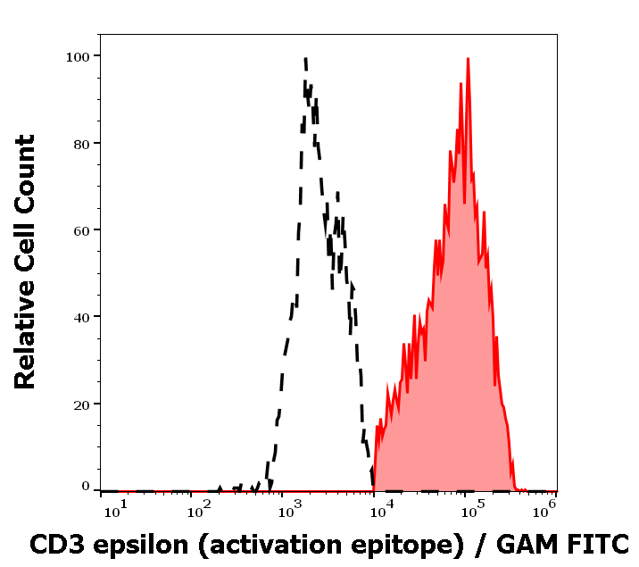
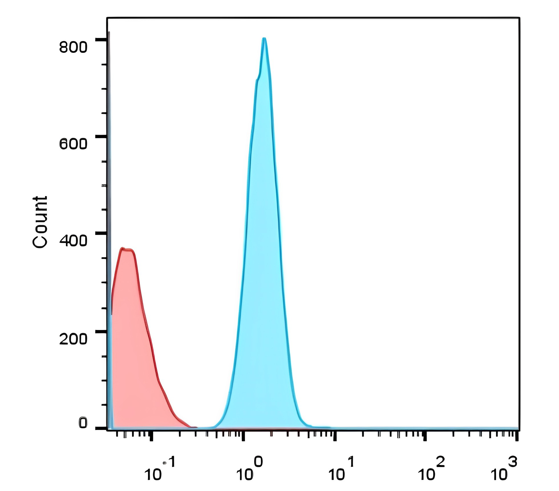
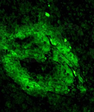
Separation of Jurkat cells stained anti-human CD3 activation epitope (APA1/1) purified antibody (concentration in sample 5 µg/ml, GAM FITC, red-filled) from Jurkat cells unstained by primary antibody (GAM FITC, black-dashed) in flow cytometry analysis (intracellular staining).
- Item 1 of 10
- Item 1 of 8
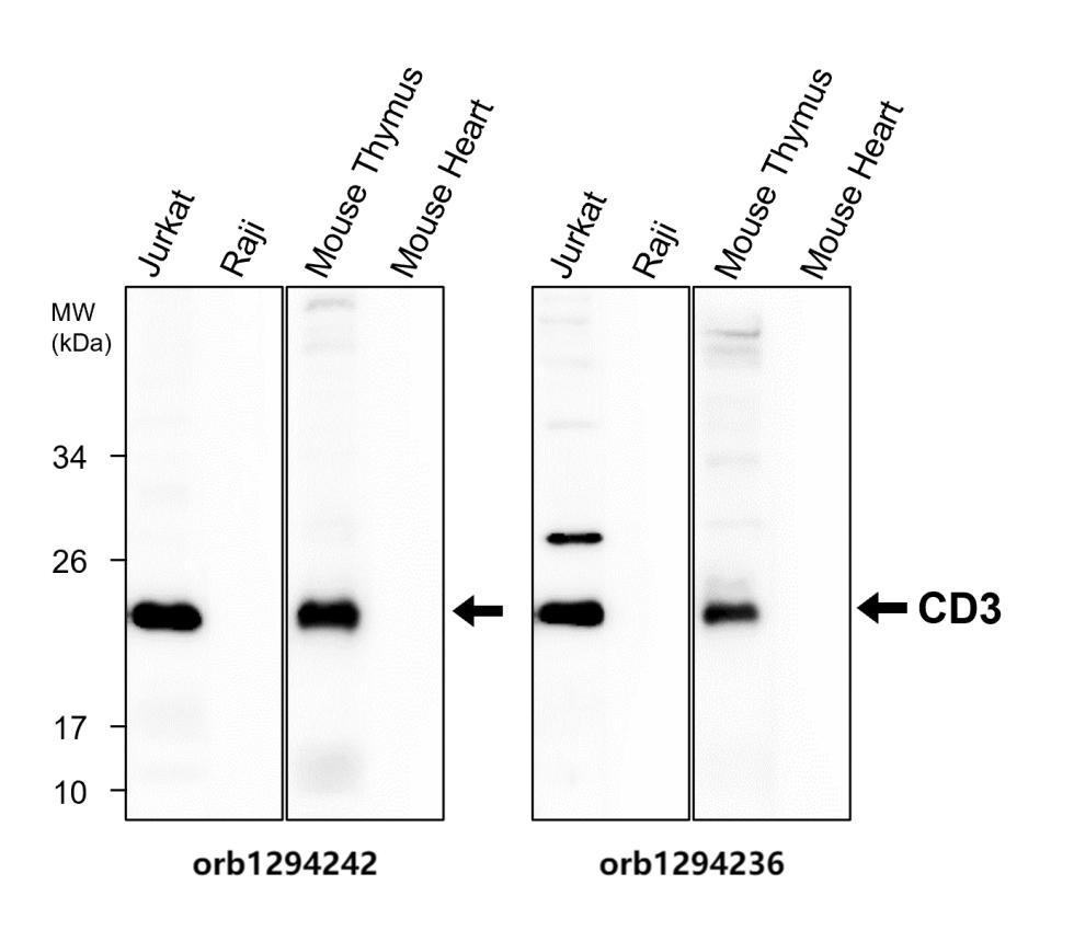
CD3 Intracellular domain Antibody [orb1294242]
IF, IHC, WB
Human, Mouse, Rabbit
Rabbit
Polyclonal
Unconjugated
100 μl, 25 μl - Item 1 of 6
- Item 1 of 5
- Item 1 of 4

Submit a review
Filter by Rating

- 5 stars

- 4 stars
- 3 stars
- 2 stars
- 1 stars