You have no items in your shopping cart.
Cart summary
Item 1 of 5
Item 1 of 5
CD20 Antibody
Catalog Number: orb2638167

| Catalog Number | orb2638167 |
|---|---|
| Category | Antibodies |
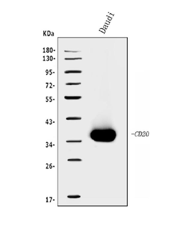
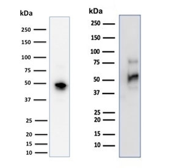
| Description | Recognizes a protein of 30-33kDa, which is identified as CD20. It is a non-Ig differentiation antigen of B-cells and its expression is restricted to normal and neoplastic B-cells, being absent from all other leukocytes and tissues. CD20 is expressed by pre B-cells and persists during all stages of B-cell maturation but is lost upon terminal differentiation into plasma cells. This MAb can be used for immunophenotyping of leukemia and malignant cells, B lymphocyte detection in peripheral blood and B cell localization in tissues. It reacts with the majority of B-cells present in peripheral blood and lymphoid tissues and their derived lymphomas. In lymphoid tissue, germinal center blasts and B-immunoblasts are particularly reactive. It is a reliable antibody for ascribing a B-cell phenotype in known lymphoid tissues. Rarely, CD20-positive T-cell lymphomas have been reported. Reactivity has also been noted with Reed-Sternberg cells in cases of Hodgkin s disease, particularly of lymphocyte predominant type. |
| Species/Host | Mouse |
| Clonality | Monoclonal |
| Clone Number | IGEL/773 |
| Tested applications | FACS, IF, IHC-P |
| Reactivity | Human |
| Isotype | Mouse IgG2a, kappa |
| Immunogen | Recombinant CD20 protein was used as the immunogen for this CD20 antibody. The epitope is localized to the cytoplasmic region of the protein. |
| Antibody Type | Primary Antibody |
| Dilution range | Flow cytometry: 1-2ug/million cells,Immunofluorescence: 1-2ug/ml,Immunohistochemistry (FFPE): 1-2ug/ml for 30 min at RT |
| Purity | Protein G affinity chromatography |
| Conjugation | Unconjugated |
| Formula | 0.2 mg/ml in 1X PBS with 0.1 mg/ml BSA (US sourced) and 0.05% sodium azide |
| Hazard Information | This CD20 antibody is available for research use only. |
| UniProt ID | P11836 |
| Storage | Maintain refrigerated at 2-8°C for up to 2 weeks. For long term storage store at -20°C in small aliquots to prevent freeze-thaw cycles. |
| Buffer/Preservatives | 0.2 mg/ml in 1X PBS with 0.1 mg/ml rAlbumin (US sourced) and 0.05% sodium azide |
| Note | For research use only |
| Application notes | The concentration stated for each application is a general starting point. Variations in protocols, secondaries and substrates may require the CD20 antibody to be titered up or down for optimal performance.1. Staining of formalin-fixed tissues requires boiling tissue sections in pH 9 10mM Tris with 1mM EDTA for 10-20 min followed by cooling at RT for 20 minutes.2. The prediluted format is supplied in a dropper bottle and is optimized for use in IHC. After epitope retrieval step (if required), drip mAb solution onto the tissue section and incubate at RT for 30 min. |
| Expiration Date | 12 months from date of receipt. |
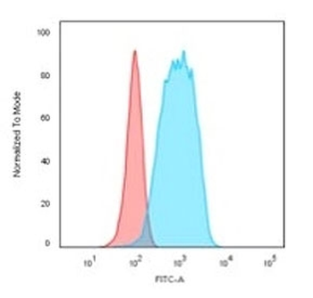
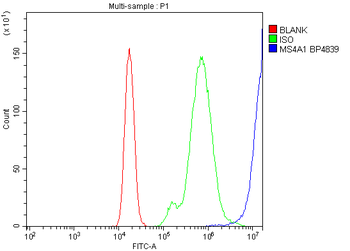
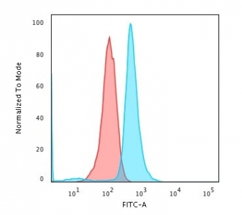
Flow cytometry testing of PFA-fixed human Raji cells with CD20 antibody (clone IGEL/773); Red = isotype control, Blue = CD20 antibody.
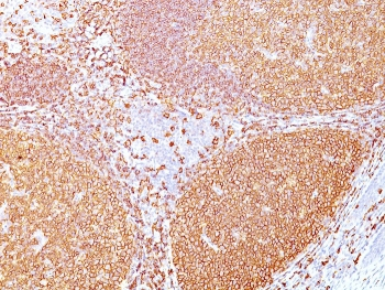
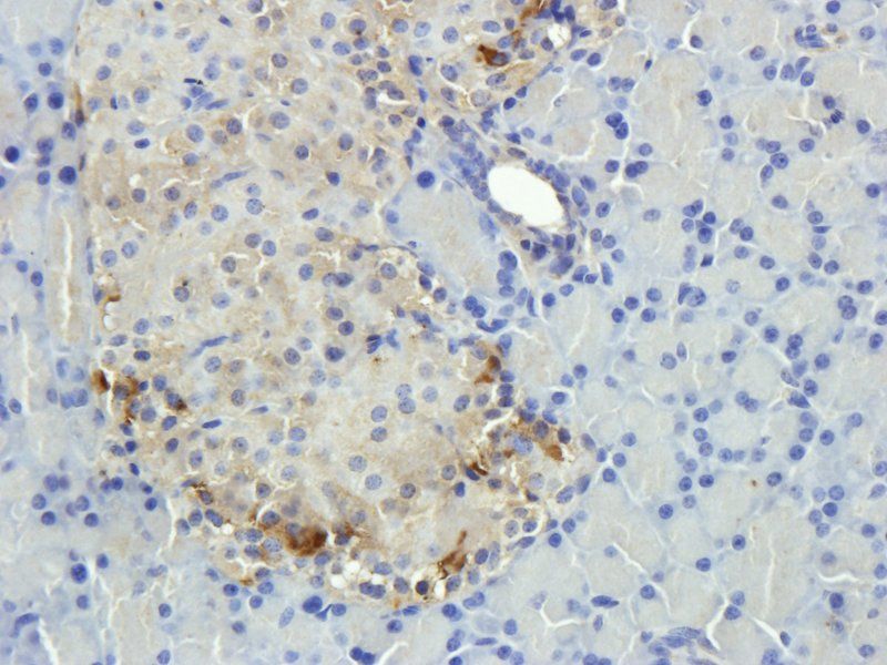
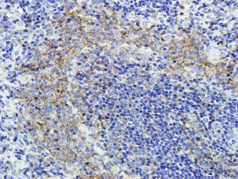
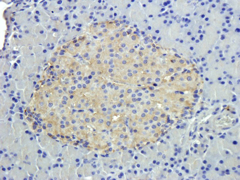
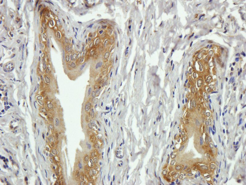
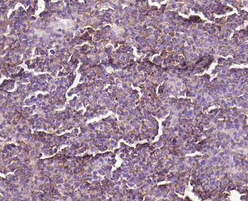
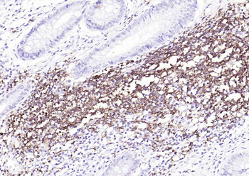
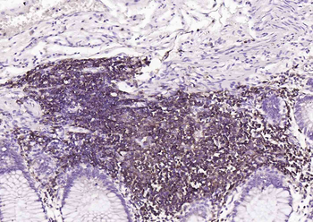
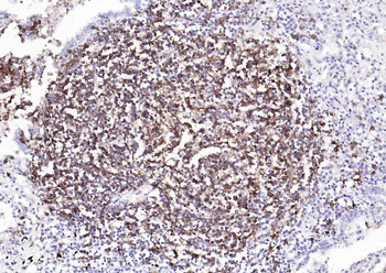
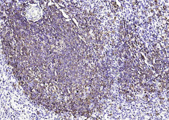
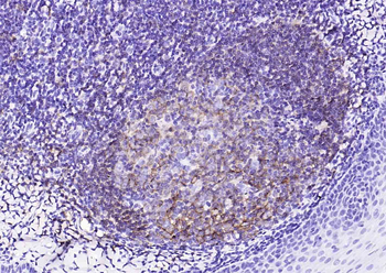
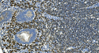
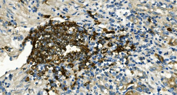
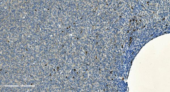
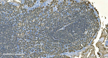
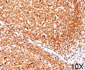
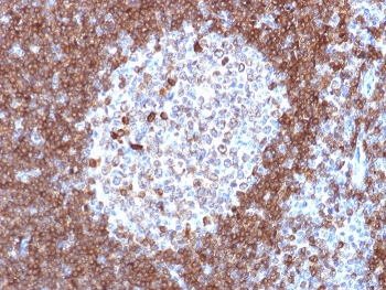
IHC staining of FFPE human tonsil tissue with CD20 antibody (clone IGEL/773).
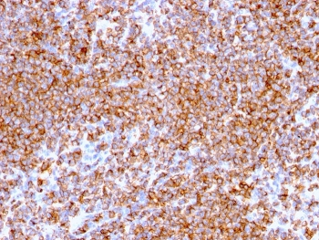
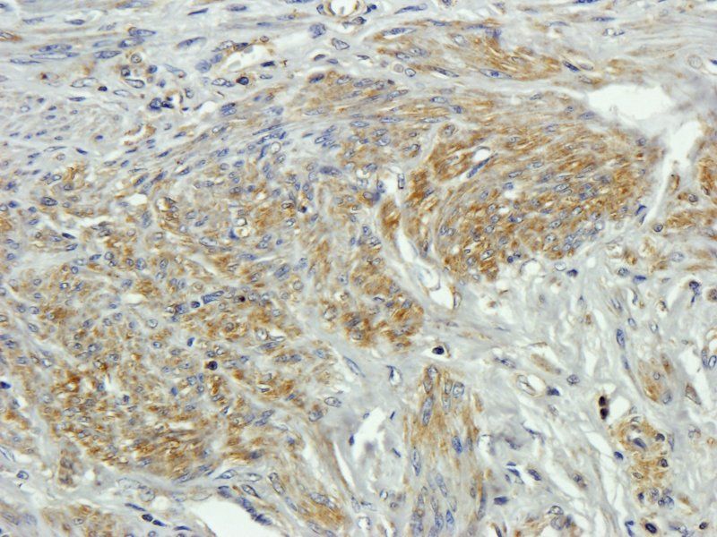
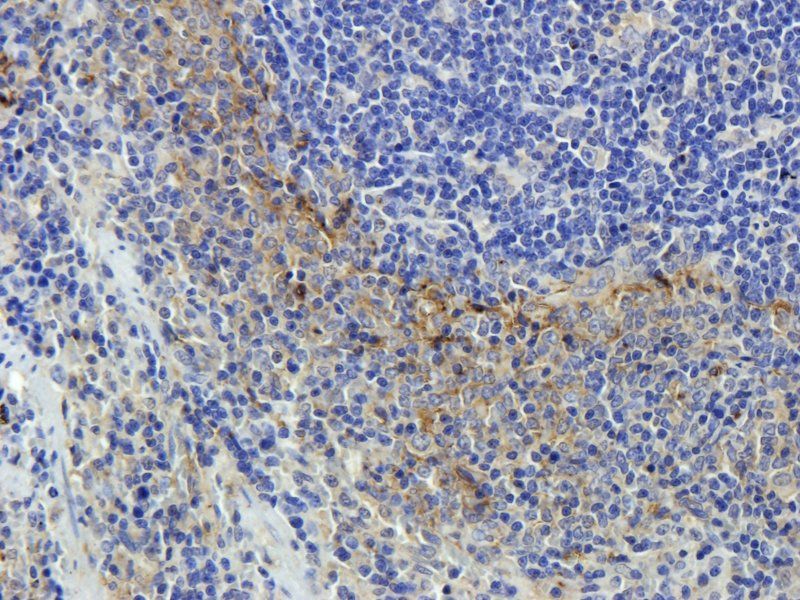
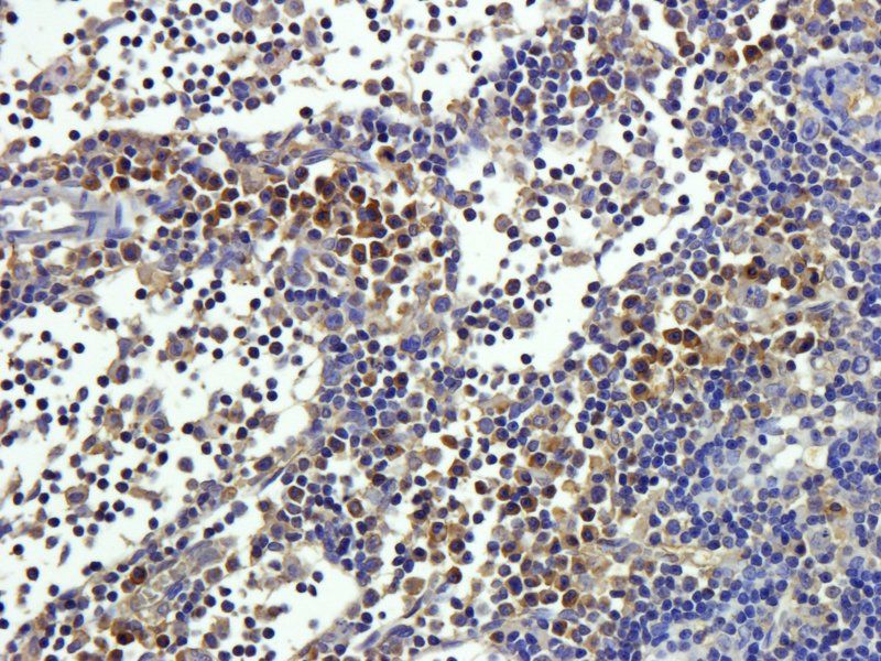
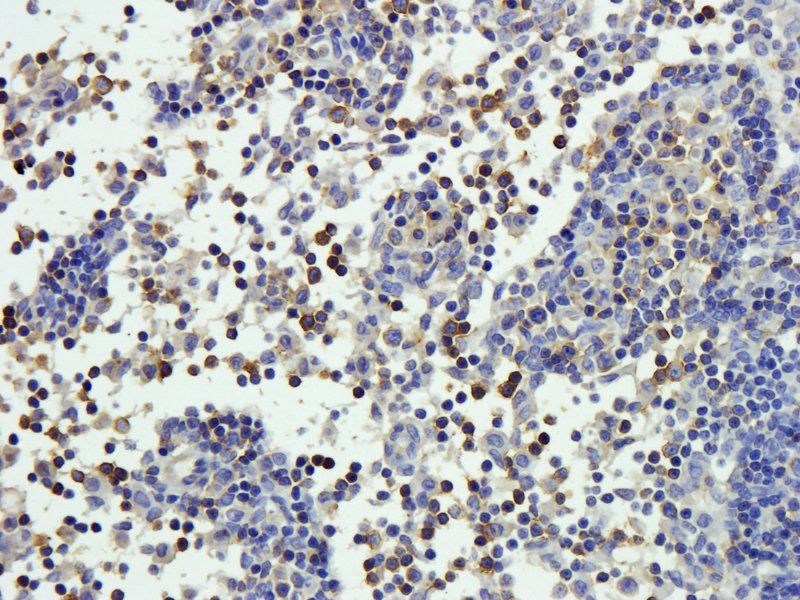
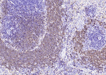
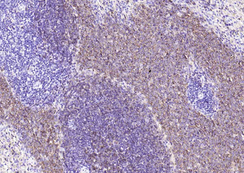
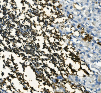
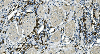
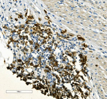
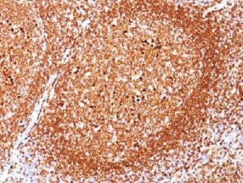
IHC staining of FFPE human lymphoma tissue with CD20 antibody (clone IGEL/773).
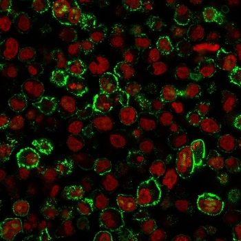
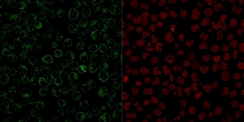
Immunofluorescent staining of PFA-fixed human Raji cells with CD20 antibody (clone IGEL/773, green) and Reddot nuclear stain (red).
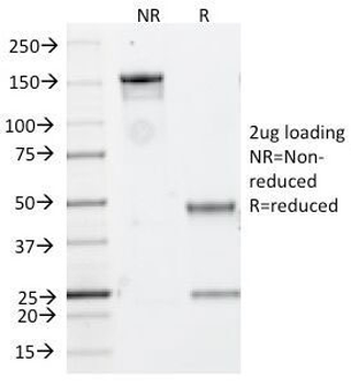
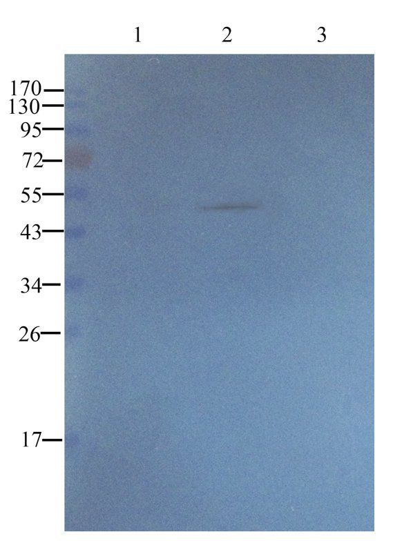
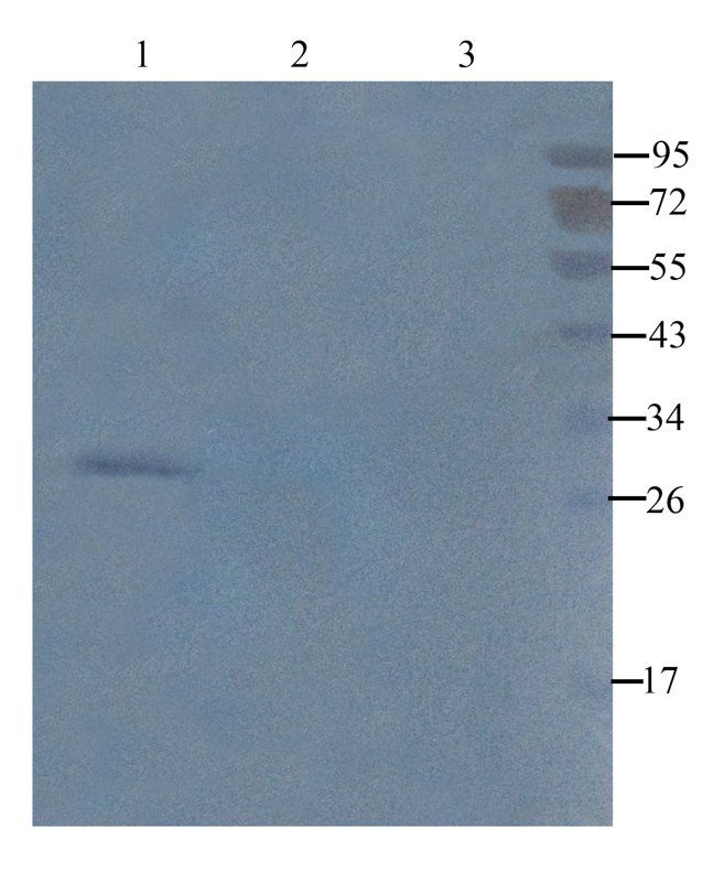
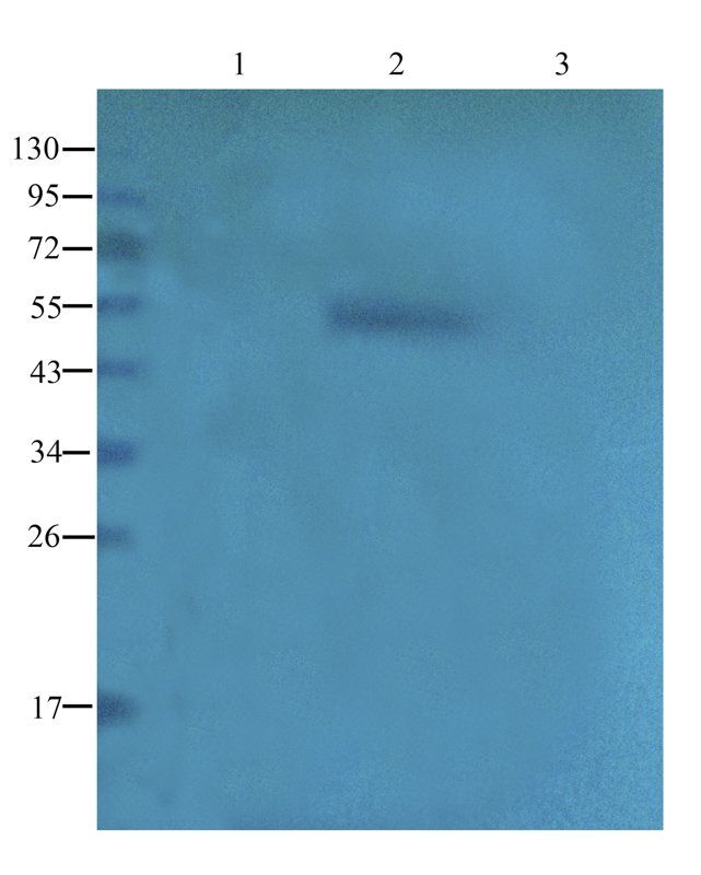
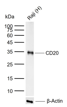
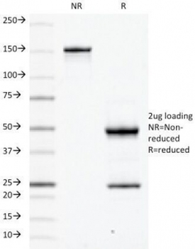
SDS-PAGE analysis of purified, BSA-free CD20 antibody (clone IGEL/773) as confirmation of integrity and purity.
- Item 1 of 11
- Item 1 of 9
CD20 Recombinant Rabbit Monoclonal Antibody [orb1499339]
IF, IHC-Fr, IHC-P, WB
Mouse, Rat
Human, Mouse, Rat
Rabbit
Recombinant
Unconjugated
100 μl, 50 μl - Item 1 of 9
Anti-CD20/MS4A1 Antibody [orb738388]
ELISA, FC, IHC, WB
Human
Rabbit
Polyclonal
Unconjugated
10 μg, 100 μg - Item 1 of 7
- Item 1 of 7