You have no items in your shopping cart.
Cart summary
Item 1 of 5
Item 1 of 5
AMACR Antibody
Catalog Number: orb1270959

| Catalog Number | orb1270959 |
|---|---|
| Category | Antibodies |
| Description | AMACR Antibody |
| Species/Host | Rabbit |
| Clonality | Polyclonal |
| Tested applications | FC, IF, IHC-P, WB |
| Reactivity | Human |
| Isotype | Rabbit Ig |
| Immunogen | This AMACR antibody is generated from rabbits immunized with a KLH conjugated synthetic peptide between 323-351 amino acids from the C-terminal region of human AMACR. |
| Antibody Type | Primary Antibody |
| Concentration | batch dependent |
| Form/Appearance | Liquid |
| Conjugation | Unconjugated |
| MW | 42 kDa |
| Target | AMACR |
| UniProt ID | Q9UHK6 |
| NCBI | Q9UHK6 |
| Storage | Maintain refrigerated at 2-8°C for up to 2 weeks. For long term storage store at -20°C in small aliquots to prevent freeze-thaw cycles. |
| Buffer/Preservatives | Supplied in PBS with 0.09% (W/V) sodium azide. |
| Alternative names | Alpha-methylacyl-CoA racemase, 2-methylacyl-CoA ra Read more... |
| Note | For research use only |
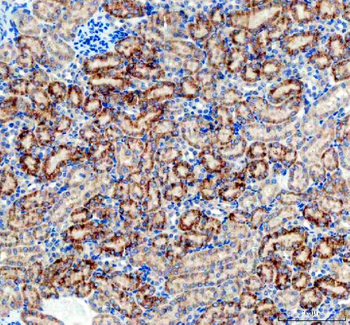
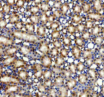
| Application notes | For WB starting dilution is: 1:1000For IHC-P starting dilution is: 1:50~100For FACS starting dilution is: 1:10~50For IF starting dilution is: 1:10~50 |
| Expiration Date | 12 months from date of receipt. |
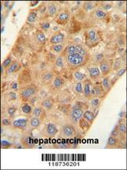
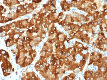
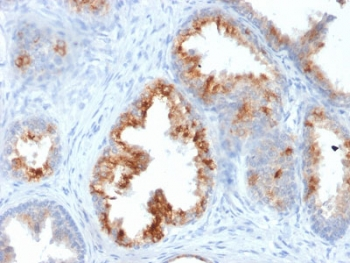
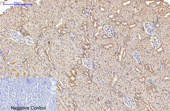
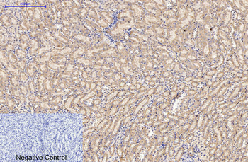
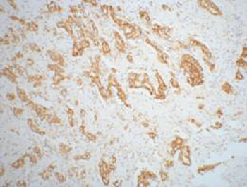
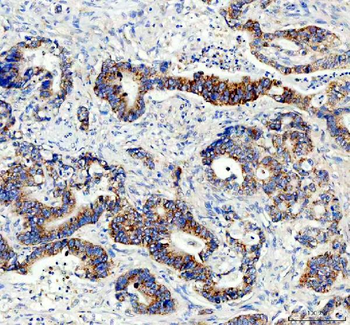
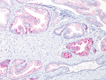
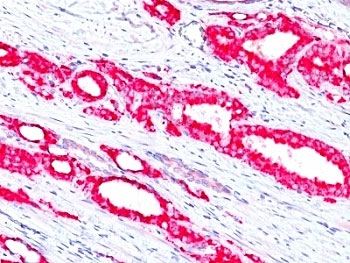
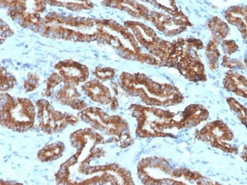
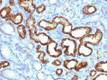
AMACR antibody immunohistochemistry analysis in formalin fixed and paraffin embedded human hepatocarcinoma followed by peroxidase conjugation of the secondary antibody and DAB staining.
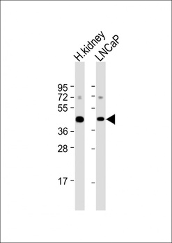
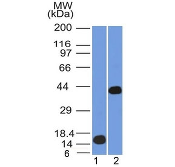
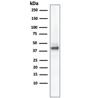
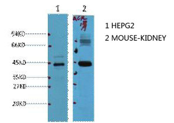
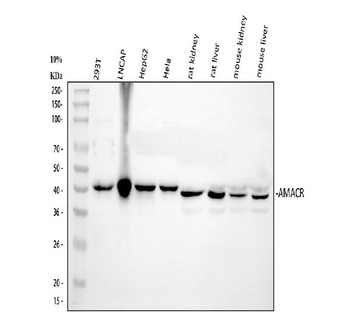
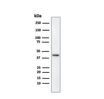
Western Blot at 1:1000 dilution Lane 1: human kidney lysate Lane 2: LNCaP whole cell lysate Lysates/proteins at 20 ug per lane.
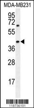
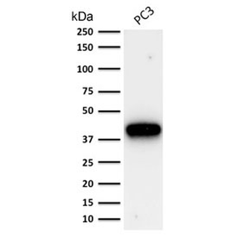
Western blot analysis in MDA-MB231 cell line lysates (35 ug/lane).
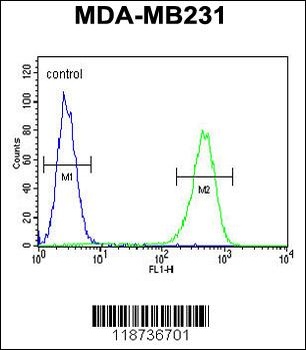
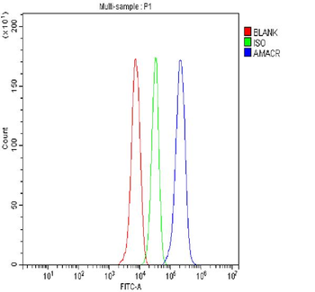
Flow cytometric analysis of MDA-MB231 cells (right histogram) compared to a negative control cell (left histogram). FITC-conjugated goat-anti-rabbit secondary antibodies were used for the analysis.
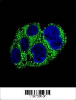
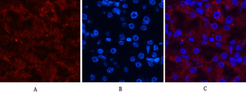
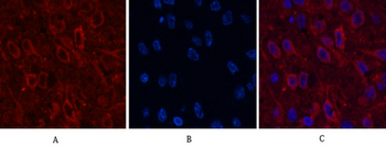
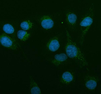
Confocal immunofluorescent analysis of AMACR Antibody with HepG2 cell followed by Alexa Fluor 488-conjugated goat anti-rabbit lgG (green). DAPI was used to stain the cell nuclear (blue).
- Item 1 of 7
AMACR / p504S Antibody (Prostate Cancer Marker) [orb606943]
IHC-P, WB
Human
Mouse
Monoclonal
Unconjugated
20 μg - Item 1 of 7
AMACR / p504S Antibody (Prostate Cancer Marker) [orb2642638]
IHC-P, WB
Human
Mouse
Monoclonal
Unconjugated
100 μg - Item 1 of 7
AMACR Monoclonal Antibody(4A12) [orb1415979]
IF, IHC-P, WB
Human, Mouse, Rat
Mouse
Monoclonal
Unconjugated
100 μl - Item 1 of 6
Anti-AMACR Antibody [orb371717]
FC, ICC, IF, IHC, WB
Human, Mouse, Rat
Rabbit
Polyclonal
Unconjugated
10 μg, 100 μg - Item 1 of 5
AMACR / p504S Antibody (Prostate Cancer Marker) [orb2638392]
IHC-P, WB
Human
Rabbit
Monoclonal
Unconjugated
0.1 ml