You have no items in your shopping cart.
Cart summary
Item 1 of 7
Item 1 of 7
AKT3
Catalog Number: orb18912

| Catalog Number | orb18912 |
|---|---|
| Category | Antibodies |
| Description | Goat polyclonal antibody to AKT3 |
| Target | AKT3 |
| Clonality | Polyclonal |
| Species/Host | Goat |
| Conjugation | Unconjugated |
| Reactivity | Bovine, Canine, Human, Mouse, Rat |
| Buffer/Preservatives | Supplied at 0.5 mg/ml in Tris saline, 0.02% sodium azide, pH 7.3 with 0.5% bovine serum albumin. Aliquot and store at -20°C. Minimize freezing and thawing. |
| Purification | Purified from goat serum by ammonium sulphate precipitation followed by antigen affinity chromatography using the immunizing peptide. |
| Protein Sequence | CSPTSQIDNIGEEEM |
| RRID | AB_10749786 |
| MW | 55.8; 54.0 |
| Tested applications | ELISA, FC, IF, IHC, WB |
| Dilution range | ELISA: 1:16000, WB: 1-3 μg/ml, IHC-P: 5 μg/ml |
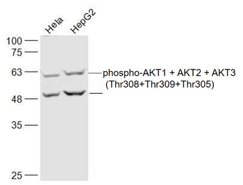
| Application notes | ELISA: Peptide ELISA: antibody detection limit dilution 1:16000.WB: A 50-55kDa band observed in Human Hepatoblastoma HepG2 lysates (calculated MW of 55.8kDa according to NP_005456 and 54.0kDa according to NP_859029). Recommended concentration: 1-3 μg/ml. |
| Storage | Aliquot and store at -20°C. Minimize freezing and thawing. |
| Alternative names | anti AKT3 antibody, anti STK-2 antibody, anti RAC- Read more... |
| Note | For research use only |
| Entrez | 10000 |
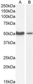
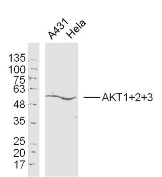
1 µg/ml staining of HepG2 (A) and HeLa (B) cell lysate (35 µg protein in RIPA buffer). Detected by chemiluminescence.
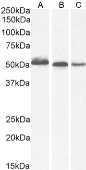
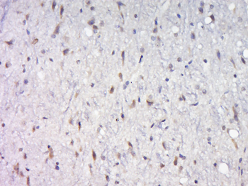
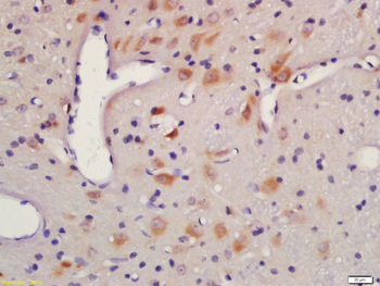
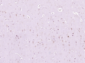
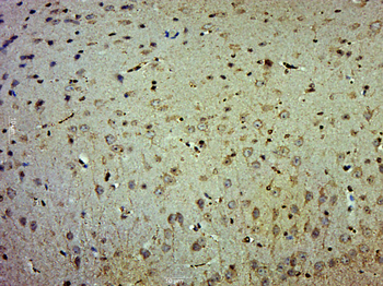
1 µg/ml staining of Human Thyroid (A), (0.5 ug/ml) Mouse Brain (B) and (1 µg/ml) Rat Brain (C) lysate (35 µg protein in RIPA buffer). Detected by chemiluminescence.
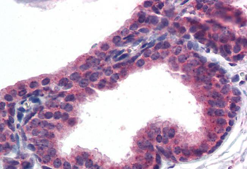
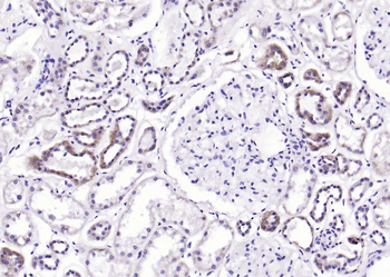
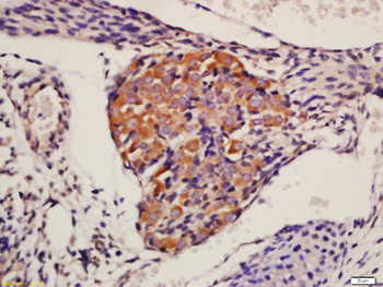
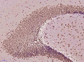
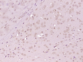
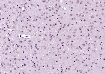
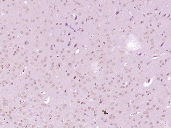
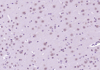
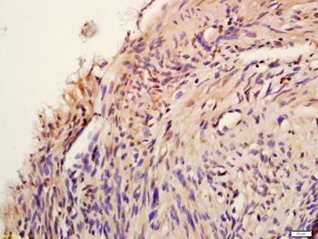
5 µg/ml staining of paraffin embedded Human Prostate. Steamed antigen retrieval with citrate buffer pH 6, AP-staining.
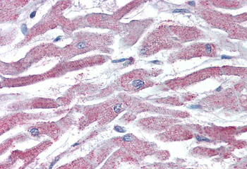
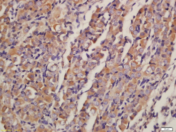
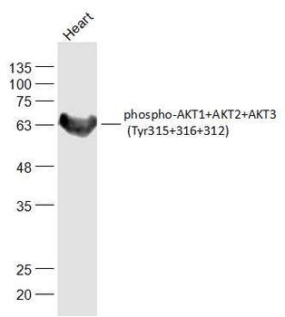
5 µg/ml staining of paraffin embedded Human Heart. Steamed antigen retrieval with citrate buffer pH 6, AP-staining.
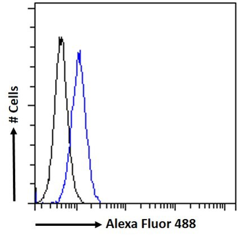
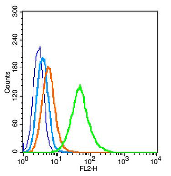
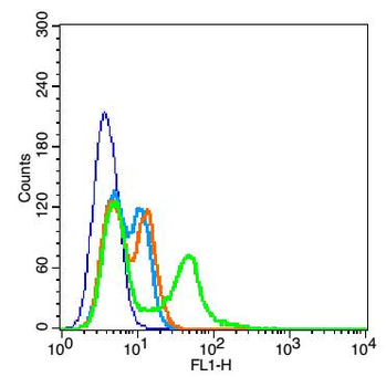
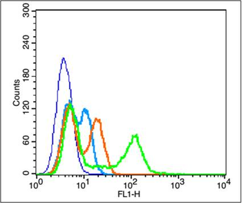
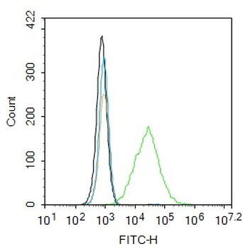
Flow cytometric analysis of paraformaldehyde fixed HepG2 cells (blue line), permeabilized with 0.5% Triton. Primary incubation 1hr (10 ug/ml) followed by Alexa Fluor 488 secondary antibody (1 ug/ml). IgG control: Unimmunized goat IgG (black line) followed by Alexa Fluor 488 secondary antibody.
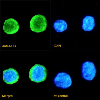
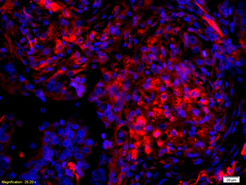
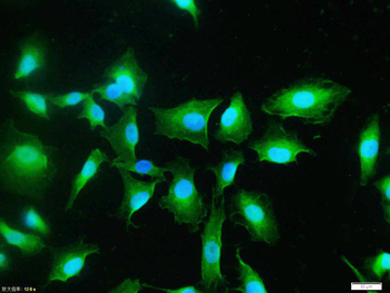
Immunofluorescence analysis of paraformaldehyde fixed THP-1 immobilized on Shi-fix™ plus cover-slips. Primary incubation 1hr (1:50 dilution) followed by Alexa Fluor® 488 secondary antibody (1:2000 dilution), showing membrane and cytoplasmic staining. The nuclear stain is DAPI (blue). Negative control: Anti-Goat IgG followed by Alexa Fluor® 488 secondary antibody.
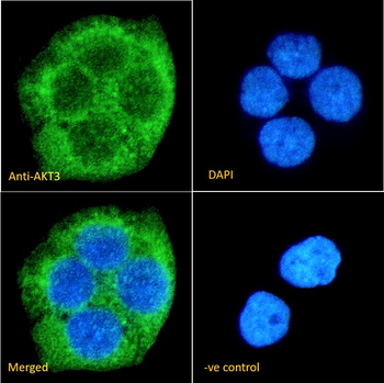
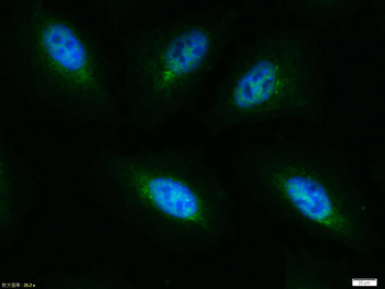
Immunofluorescence analysis of paraformaldehyde fixed A431. Primary incubation 1hr (1:50 dilution) followed by Alexa Fluor® 488 secondary antibody (1:2000 dilution), showing cytoplasmic staining. The nuclear stain is DAPI (blue). Negative control: Anti-Goat IgG followed by Alexa Fluor® 488 secondary antibody.
- Item 1 of 8
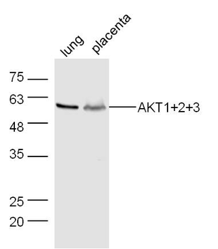
AKT1+2+3 Rabbit Polyclonal Antibody [orb155629]
FC, IF, IHC-Fr, IHC-P, WB
Bovine, Canine, Gallus, Porcine, Rabbit, Sheep
Human, Mouse, Rat
Rabbit
Polyclonal
Unconjugated
100 μl, 200 μl, 50 μl - Item 1 of 7
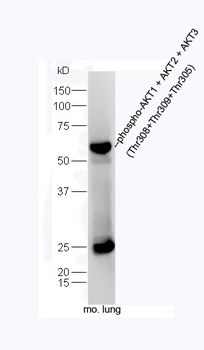
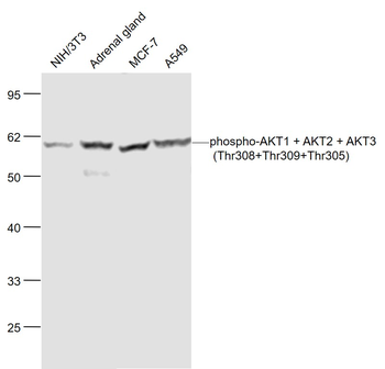
Phospho-AKT1 + AKT2 + AKT3 (Thr308+Thr309+Thr305) Rabbit Polyclonal Antibody [orb6780]
FC, ICC, IF, IHC-Fr, IHC-P, WB
Bovine, Canine, Gallus, Porcine, Rabbit, Sheep
Human, Mouse, Rat
Rabbit
Polyclonal
Unconjugated
100 μl, 50 μl, 200 μl - Item 1 of 6
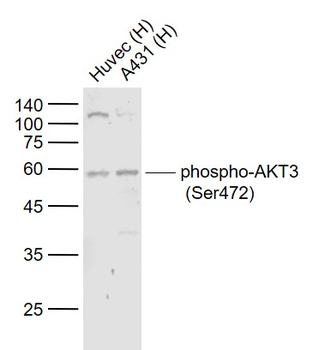
Phospho-AKT3 (Ser472) Rabbit Polyclonal Antibody [orb6790]
IF, IHC-Fr, IHC-P, WB
Bovine, Canine, Gallus, Porcine, Rabbit, Sheep
Human, Mouse, Rat
Rabbit
Polyclonal
Unconjugated
200 μl, 50 μl, 100 μl - Item 1 of 5
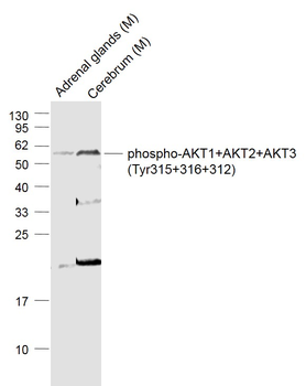
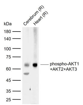
Phospho-AKT1+AKT2+AKT3 (Tyr315+316+312) Rabbit Polyclonal Antibody [orb6789]
FC, IF, IHC-Fr, IHC-P, WB
Bovine, Canine, Gallus, Porcine, Rabbit, Sheep
Human, Mouse, Rat
Rabbit
Polyclonal
Unconjugated
100 μl, 200 μl, 50 μl - Item 1 of 6
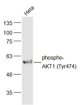
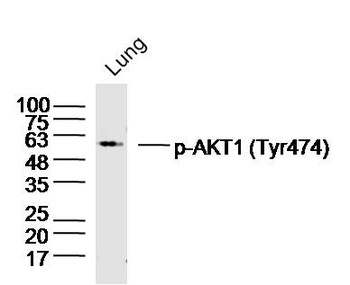
Phospho-AKT1 (Tyr474) Rabbit Polyclonal Antibody [orb155627]
FC, ICC, IF, IHC-Fr, IHC-P, WB
Bovine, Canine, Gallus, Porcine, Rabbit, Rat, Sheep
Human, Mouse
Rabbit
Polyclonal
Unconjugated
50 μl, 100 μl, 200 μl