You have no items in your shopping cart.
Cart summary
Item 1 of 5
Item 1 of 5
AK4 Antibody (Center)
Catalog Number: orb1788442

| Catalog Number | orb1788442 |
|---|---|
| Category | Antibodies |
| Description | Purified Rabbit Polyclonal Antibody (Pab) |
| Target | This AK4 antibody is generated from a rabbit immunized with a KLH conjugated synthetic peptide between 119-153 amino acids from the Central region of human AK4. |
| Clonality | Polyclonal |
| Species/Host | Rabbit |
| Isotype | Rabbit IgG |
| Conjugation | Unconjugated |
| Reactivity | Human |
| Predicted Reactivity | Mouse, Rat |
| Form/Appearance | Purified polyclonal antibody supplied in PBS with 0.09% (W/V) sodium azide. This antibody is purified through a protein A column, followed by peptide affinity purification. |
| Immunogen | 119-153 aa |
| UniProt ID | P27144 |
| MW | 25268 Da |
| Tested applications | IF, IHC-P, WB |
| Dilution range | IF: 1:25, WB: 1:1000, IHC-P: 1:25, IHC-P: 1:25, IHC-P: 1:25 |
| Antibody Type | Primary Antibody |
| Storage | Maintain refrigerated at 2-8°C for up to 2 weeks. For long term storage store at -20°C in small aliquots to prevent freeze-thaw cycles |
| Alternative names | Adenylate kinase 4, mitochondrial, AK 4, Adenylate Read more... |
| Research Area | Cell Biology |
| Note | For research use only |
| Expiration Date | 12 months from date of receipt. |
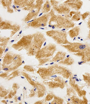
Immunohistochemical analysis of paraffin-embedded H.heart section using AK4 Antibody (Center). Diluted at 1:25 dilution. A peroxidase-conjugated goat anti-rabbit IgG at 1:400 dilution was used as the secondary antibody, followed by DAB staining.

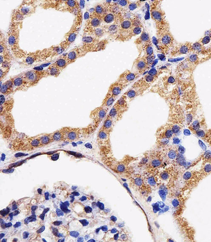
Immunohistochemical analysis of paraffin-embedded H.kidney section using AK4 Antibody (Center). Diluted at 1:25 dilution. A peroxidase-conjugated goat anti-rabbit IgG at 1:400 dilution was used as the secondary antibody, followed by DAB staining.
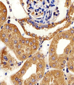
Immunohistochemical analysis of paraffin-embedded M.kidney section using AK4 Antibody (Center). Diluted at 1:25 dilution. A peroxidase-conjugated goat anti-rabbit IgG at 1:400 dilution was used as the secondary antibody, followed by DAB staining.
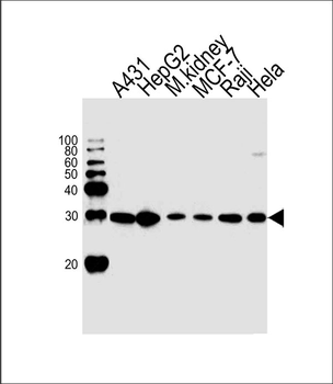
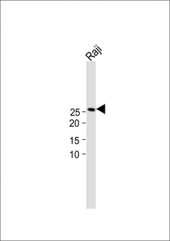
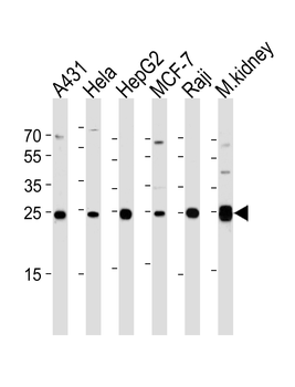
Western blot analysis of lysates from A431, HepG2 cell line, mouse kidney tissue, MCF-7, Raji, Hela cell line (from left to right), using AK4 Antibody (Center). Diluted at 1:1000 at each lane. A goat anti-rabbit IgG H&L (HRP) at 1:10000 dilution was used as the secondary antibody.
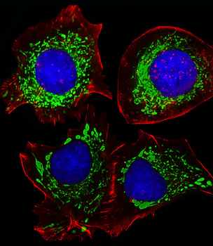
Fluorescent image of HepG2 cells stained with AK4 Antibody (Center). Diluted at 1:25 dilution. An Alexa Fluor 488-conjugated goat anti-rabbit lgG at 1:400 dilution was used as the secondary antibody (green). DAPI was used to stain the cell nuclear (blue). Cytoplasmic actin was counterstained with Alexa Fluor 555 conjugated with Phalloidin (red).
- Item 1 of 6
AK4 Antibody (Center) [orb1927188]
IF, IHC-P, WB
Human, Mouse
Rabbit
Polyclonal
Unconjugated
100 μl, 50 μl