You have no items in your shopping cart.
Cart summary
Item 1 of 11
Item 1 of 11
AIFM1 Antibody
Catalog Number: orb1239169
| Catalog Number | orb1239169 |
|---|---|
| Category | Antibodies |
| Description | AIFM1 Antibody |
| Species/Host | Rabbit |
| Clonality | Polyclonal |
| Tested applications | ELISA, IHC-P, WB |
| Reactivity | Human, Mouse, Rat |
| Isotype | IgG |
| Immunogen | Anti-AIF antibody (orb1239169) was raised against a peptide corresponding to 15 amino acids near the center of human AIF. The immunogen is located within amino acids 500-550 of AIF. |
| Concentration | 1 mg/mL |
| Dilution range | WB: 1-2 μg/mL; IHC-P: 10 μg/mL.Antibody validated: Western Blot in human, mouse, and rat samples; Immunohistochemistry in human samples. All other applications and species not yet tested. |
| Form/Appearance | Liquid |
| Conjugation | Unconjugated |
| MW | Predicted: 67 kDa Observed: 68 kDa |
| Target | AIFM1 |
| UniProt ID | O95381 |
| NCBI | O95381 |
| Storage | AIF antibody can be stored at 4°C for three months and -20°C, stable for up to one year. As with all antibodies care should be taken to avoid repeated freeze thaw cycles. Antibodies should not be exposed to prolonged high temperatures. |
| Buffer/Preservatives | AIF Antibody is supplied in PBS containing 0.02% sodium azide. |
| Alternative names | AIF Antibody: Apoptosis-inducing factor 1, Program Read more... |
| Note | For research use only |
| Expiration Date | 12 months from date of receipt. |
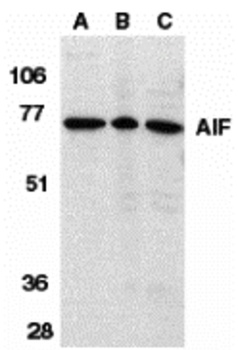
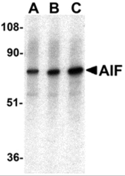
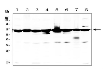
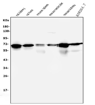
Western Blot Validation in Different Species. Loading: 15 µg of lysates per lane. Antibodies: AIF orb1239169, (1 µg/mL), 1h incubation at RT in 5% NFDM/TBST. Secondary: Goat anti-rabbit IgG HRP conjugate at 1:10000 dilution. Lane A: Human K562 cells, Lane B: Rat heart, Lane C: Mouse heart.
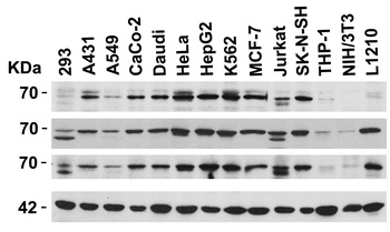
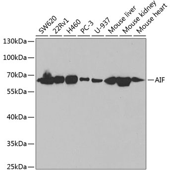
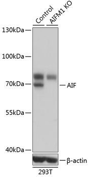
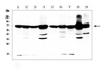
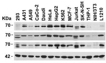
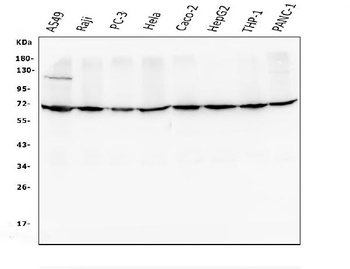
Independent Antibody Validation (IAV) via Protein Expression Profile in Cell Lines. Loading: 15 µg of lysates per lane. Antibodies: AIF orb1239169, (1 µg/mL), AIF orb1239191, (1 µg/mL), AIF orb1239168, (2 µg/mL), and beta-actin (1 µg/mL), 1h incubation at RT in 5% NFDM/TBST. Secondary: Goat anti-rabbit IgG HRP conjugate at 1:10000 dilution.
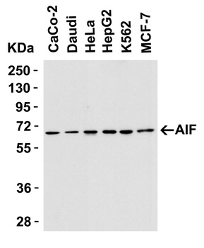
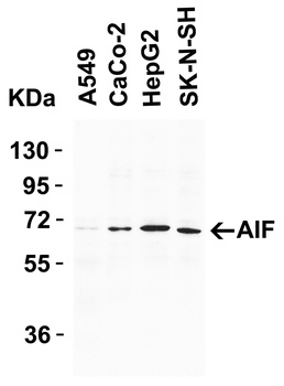
Western Blot Validation in Human Cell Lines. Loading: 15 µg of lysates per lane. Antibodies: AIF orb1239169, (1 µg/mL), 1h incubation at RT in 5% NFDM/TBST. Secondary: Goat anti-rabbit IgG HRP conjugate at 1:10000 dilution.
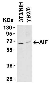
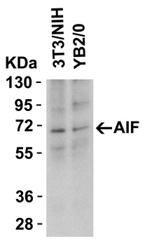
Western Blot Validation in Mouse and Rat Cell Lines. Loading: 15 µg of lysates per lane. Antibodies: AIF orb1239169, (1 µg/mL), 1h incubation at RT in 5% NFDM/TBST. Secondary: Goat anti-rabbit IgG HRP conjugate at 1:10000 dilution.
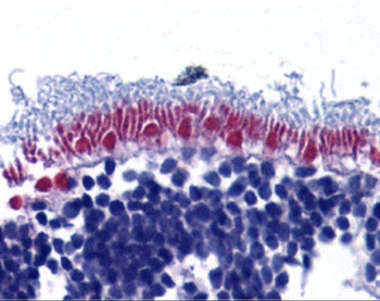
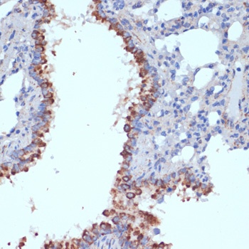
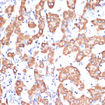
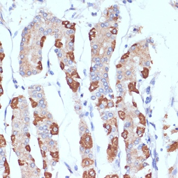
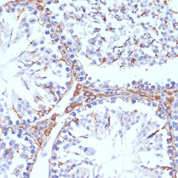
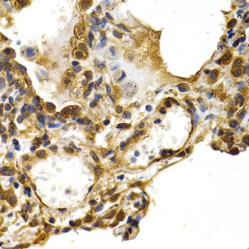
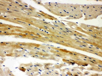
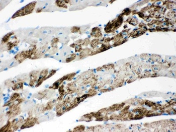
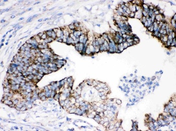
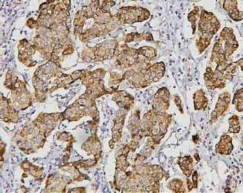
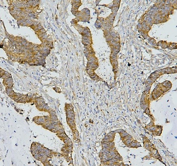
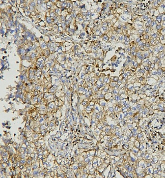
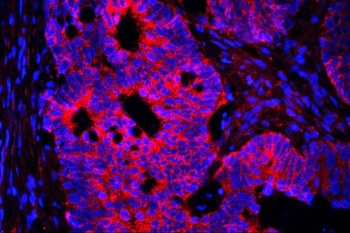
Immunohistochemistry Validation of AIF in Human Retina Tissue. Immunohistochemical analysis of paraffin-embedded human retina tissue using anti-AIF antibody (orb1239169) at 10 µg/ml. Tissue was fixed with formaldehyde and blocked with 10% serum for 1 h at RT; antigen retrieval was by heat mediation with a citrate buffer (pH6). Samples were incubated with primary antibody overnight at 4°C. A goat anti-rabbit IgG H&L (HRP) at 1/250 was used as secondary. Counter stained with Hematoxylin.
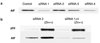
KD and Induced Validation of AIF in H1299 Cells (Stambolsky et al., 2006). Western blot analysis of AIF knockdown with anti-AIF antibodies in H1299 cells. AIF expression was disrupted in AIF knockdown cells (siRNA1 and siRNA4). An increased expression of AIF was induced by ZnCl2 treatment, which was not observed in AIF knockdown cells.

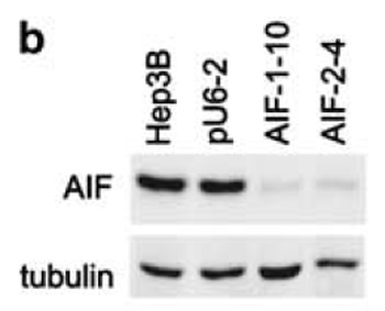
KD Validation of AIF in AIF Silenced Stable Cells (Apostolova et al., 2006). AIF silencing is sustained in stable cell lines. Western blot analysis ofstable lines AIF-1-10, AIF-2-4 and pU6-2 using anti-AIF antibodies. AIF protein was disrupted after AIF silencing with AIF siRNA (AIF-1-10 and AIF-2-4) as compared to control (Hep3B and pU6-2).
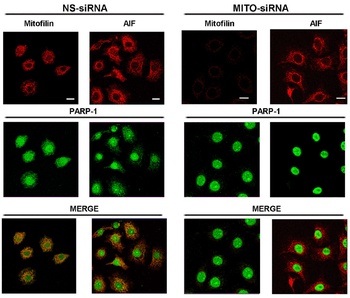
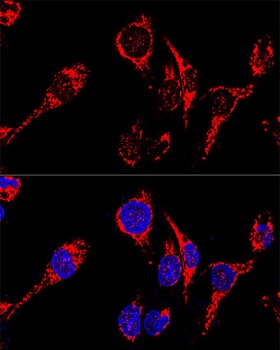
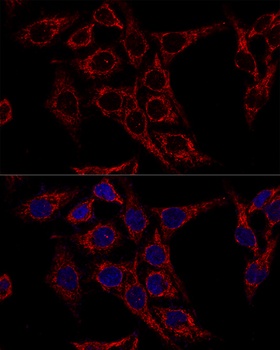
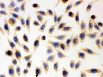
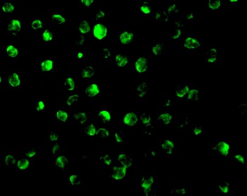
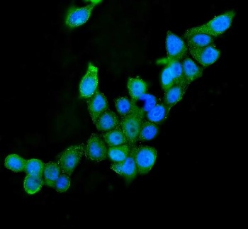
Immunofluorescence Validation of AIF in HeLa Cells (Rossi et al., 2009). HeLa cells were transfected withMitofilin-specific (MITO-siRNA) or with nonspecific (NS-siRNA) siRNAs. AIF staining with anti-AIF antibodies was shown as a mitochondrial marker in the absence of Mitofilin.

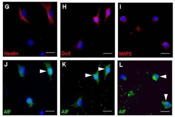
Immunofluorescence Validation of AIF in Rat Hippocampal Neurons (Hofer et al., 2011). (G-L) After exposure to bacterial components, AIF colocalized in mature neurons (MAP2; I, L), immature neurons (DcX; H, K), and stem/progenitor cells (Nestin; G, J). AIF expression was detected by anti-AIF antibodies.
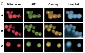
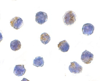
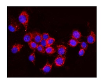
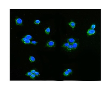
Subcellular Localization Validation of AIF in mononuclear cells (Gupta et al., 2003). A shows mononuclear cells (MNCs) alone, B shows MNCs transfected with control plasmid, C shows MNCs transfected with Bcl-2 expression plasmid. Overlay is of Mitotracker (red) and AIF (green). Hoechst 33258 dye is used to examine chromatin fragmentation. The release of AIF form mitochondria is detected by anti-AIF antibodies.

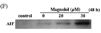
Induced Expression Validation of AIF in U937 Cells (Ikai et al., 2006). Release of AIF at 48 h after the treatment with 30 uM magnolol examined by Western blotAnalysis with anti-AIF antibodies. AIF release was markedly increased 48h after magnolol treatment.
- Item 1 of 10
- Item 1 of 9
- Item 1 of 9
AIF/AIFM1 Antibody [orb251549]
FC, ICC, IF, IHC, WB
Hamster
Human, Mouse, Rat
Rabbit
Polyclonal
Unconjugated
10 μg, 100 μg - Item 1 of 8
AIFM1 Antibody [orb1239191]
ELISA, IF, WB
Mouse, Rat
Human
Rabbit
Polyclonal
Unconjugated
0.1 mg, 0.02 mg - Item 1 of 8
AIF/AIFM1 Antibody (monoclonal, 2I5) [orb547790]
FC, ICC, IF, IHC, WB
Human, Mouse, Rat
Mouse
Monoclonal
Unconjugated
10 μg, 100 μg


Submit a review
Filter by Rating

- 5 stars

- 4 stars
- 3 stars
- 2 stars
- 1 stars