You have no items in your shopping cart.
Cart summary
Item 1 of 4
Item 1 of 4
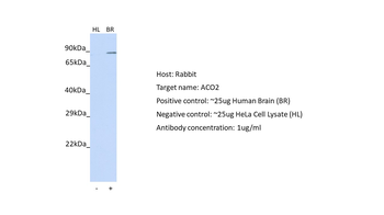
ACO2 Antibody
Catalog Number: orb1265557

| Catalog Number | orb1265557 |
|---|---|
| Category | Antibodies |
| Description | ACO2 Antibody |
| Species/Host | Rabbit |
| Clonality | Polyclonal |
| Tested applications | IHC-P, WB |
| Predicted Reactivity | Bovine, Porcine |
| Reactivity | Human, Rat |
| Isotype | Rabbit Ig |
| Immunogen | This ACO2 antibody is generated from rabbits immunized with a KLH conjugated synthetic peptide between 438-467 amino acids from the Central region of human ACO2. |
| Antibody Type | Primary Antibody |
| Concentration | batch dependent |
| Form/Appearance | Liquid |
| Conjugation | Unconjugated |
| MW | 85 kDa |
| Target | ACO2 |
| UniProt ID | Q99798 |
| NCBI | Q99798 |
| Storage | Maintain refrigerated at 2-8°C for up to 2 weeks. For long term storage store at -20°C in small aliquots to prevent freeze-thaw cycles. |
| Buffer/Preservatives | Supplied in PBS with 0.09% (W/V) sodium azide. |
| Alternative names | Aconitate hydratase, mitochondrial, Aconitase, Cit Read more... |
| Note | For research use only |
| Application notes | For WB starting dilution is: 1:1000For IHC-P starting dilution is: 1:10~50 |
| Expiration Date | 12 months from date of receipt. |
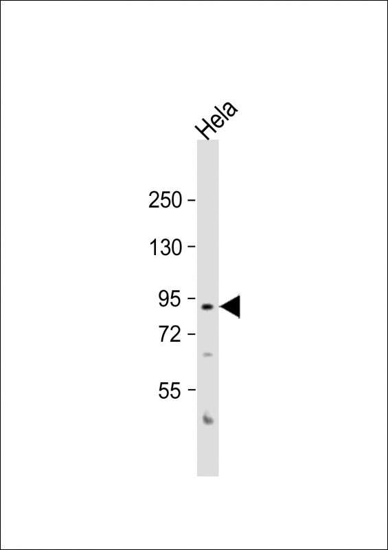
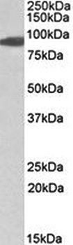
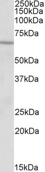
Western Blot at 1:1000 dilution + Hela whole cell lysate Lysates/proteins at 20 ug per lane.
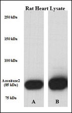
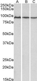
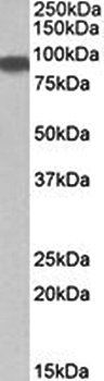
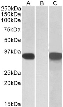
Perfused isolated rat heart whole tissue lysate was lysed with either A) 50 mM Tris-HCl, 150 mM NaCl, 1 mM EDTA, 1% NP-40, 0.1% SDS, 0.5% Na-deoxycholate, 1 mM Na3VO4, 20 mM NaF, 1 mM PMSF, 5 v/v % protease inhibitor cocktail or B) T-PER Tissue Protein Extraction Reagent, containing 1mM Na3VO4, 20 mM NaF, 5 v/v % protease inhibitor cocktail (Sigma) ; PVDF membrane was incubated in primary Ab [rabbit polyclonal antibody against ACO2 (Center). Solution: 1:1000 diluted in 5% NFM TBS-T 0, 05 for overnight (15 hrs) at 4 ?.
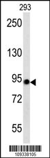
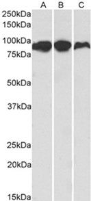
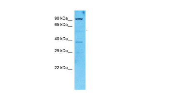
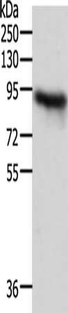
Western blot analysis in 293 cell line lysates (35 ug/lane). This demonstrates the Aconitase antibody detected the Aconitase protein (arrow).
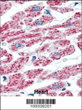
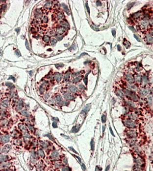
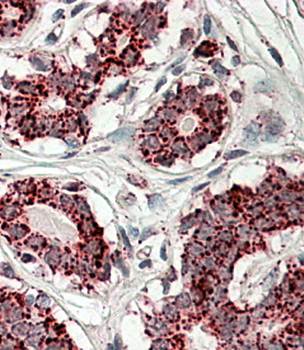
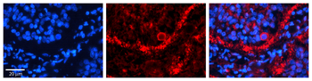
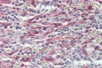
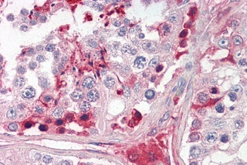
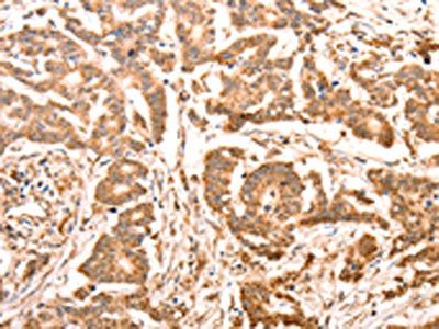
Formalin-fixed and paraffin-embedded human Heart tissue reacted with ACO2 Antibody, which was peroxidase-conjugated to the secondary antibody, followed by AEC staining.
- Item 1 of 4
Goat anti-Aconitase 2 (aa541-555) Antibody [orb20461]
ELISA, IHC, WB
Bovine, Canine, Human, Mouse, Porcine, Rat
Goat
Polyclonal
Unconjugated
100 μg - Item 1 of 2
Goat anti-Aconitase 2 Antibody [orb20462]
ELISA, IF, IHC, WB
Bovine, Canine, Human, Mouse, Porcine, Rat
Goat
Polyclonal
Unconjugated
100 μg - Item 1 of 4
ACO2 Rabbit Polyclonal Antibody [orb330838]
IHC, WB
Bovine, Porcine, Rat, Yeast
Human
Rabbit
Polyclonal
Unconjugated
100 μl - Item 1 of 4
ACO2 Antibody [orb1249162]
ELISA, IHC, WB
Bovine, Canine
Human, Mouse, Porcine, Rat
Goat
Polyclonal
Unconjugated
0.1 mg - Item 1 of 3