You have no items in your shopping cart.
Cart summary
Item 1 of 6
Item 1 of 6
XRCC6 Antibody
Catalog Number: orb1264324

| Catalog Number | orb1264324 |
|---|---|
| Category | Antibodies |
| Description | XRCC6 Antibody |
| Target | XRCC6 |
| Clonality | Polyclonal |
| Isotype | Rabbit Ig |
| Conjugation | Unconjugated |
| Reactivity | Human |
| Form/Appearance | Liquid |
| Concentration | batch dependent |
| Buffer/Preservatives | Supplied in PBS with 0.09% (W/V) sodium azide. |
| Purification | This antibody is purified through a protein A column, followed by peptide affinity purification. |
| Immunogen | This XRCC6 antibody is generated from rabbits immunized with a KLH conjugated synthetic peptide between 521-548 amino acids from the C-terminal region of human XRCC6. |
| UniProt ID | P12956 |
| MW | 70 kDa |
| Tested applications | FC, IF, IHC-P, WB |
| Application notes | For WB starting dilution is: 1:1000For IHC-P starting dilution is: 1:50~100For FACS starting dilution is: 1:10~50For IF starting dilution is: 1:10~50 |
| Antibody Type | Primary Antibody |
| Storage | Maintain refrigerated at 2-8°C for up to 2 weeks. For long term storage store at -20°C in small aliquots to prevent freeze-thaw cycles. |
| Alternative names | X-ray repair cross-complementing protein 6, 364-, Read more... |
| Note | For research use only |
| NCBI | P12956 |
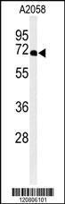
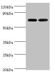
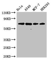
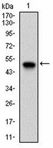
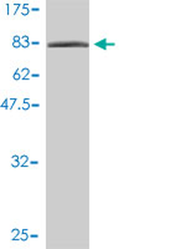
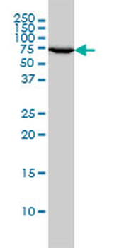
Western blot analysis of XRCC6 Antibody in A2058 cell line lysates (35 ug/lane)
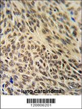
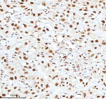
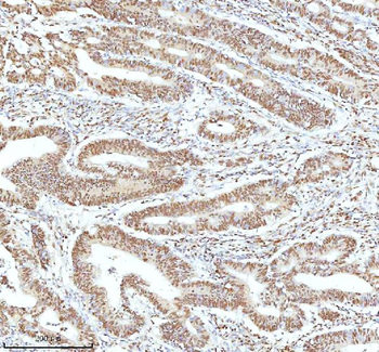
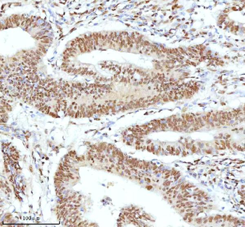
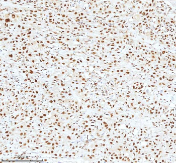
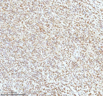
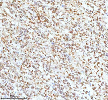
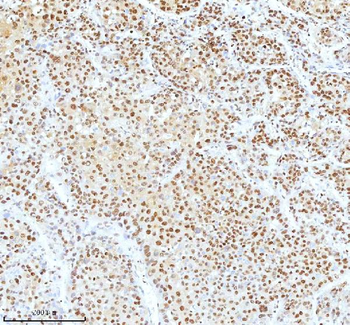
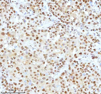
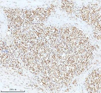
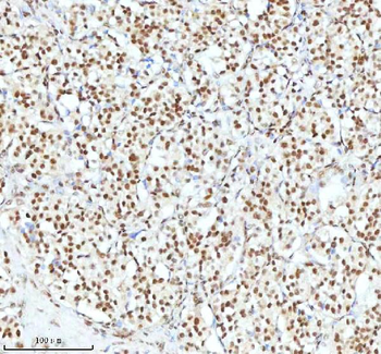
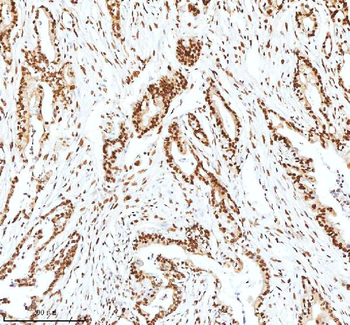
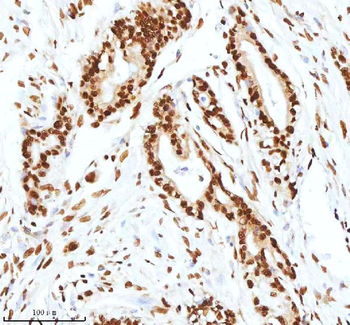
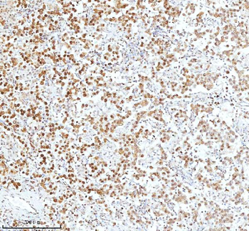
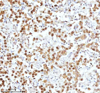
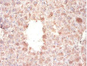
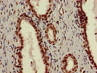
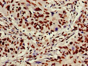
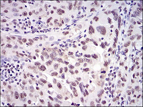
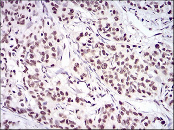
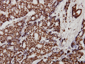
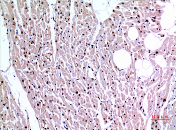
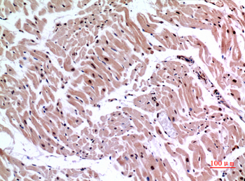
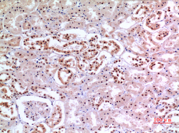
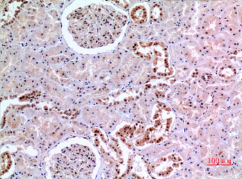
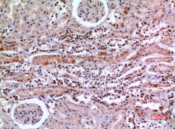
XRCC6 Antibody IHC analysis in formalin fixed and paraffin embedded human lung carcinoma followed by peroxidase conjugation of the secondary antibody and DAB staining.

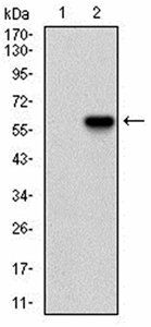
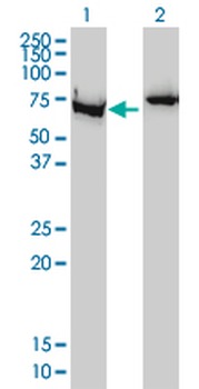
Western blot analysis of XRCC6 using rabbit polyclonal XRCC6 Antibody using 293 cell lysates (2 ug/lane) either nontransfected (Lane 1) or transiently transfected (Lane 2) with the XRCC6 gene.
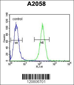
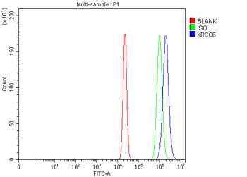
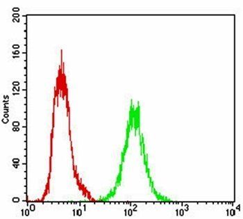
Flow cytometric analysis of A2058 cells (right histogram) compared to a negative control cell (left histogram). FITC-conjugated goat-anti-rabbit secondary antibodies were used for the analysis.
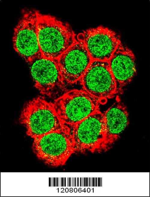
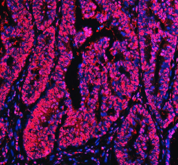
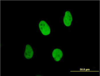
Confocal immunofluorescent analysis of XRCC6 Antibody with 293 cell followed by Alexa Fluor 488-conjugated goat anti-rabbit lgG (green). Actin filaments have been labeled with Alexa Fluor 555 phalloidin (red).
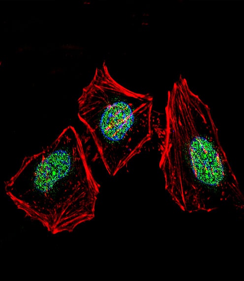
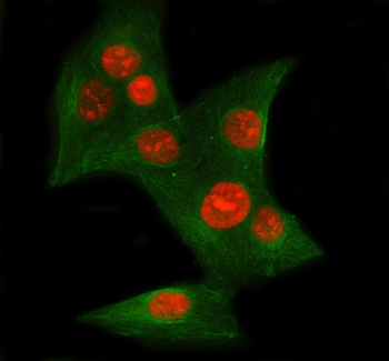
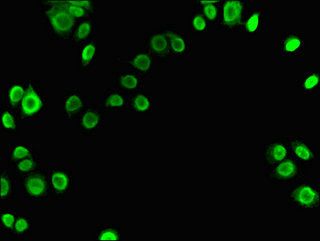
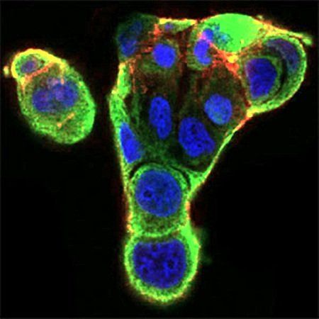
Fluorescent confocal image of Hela cell stained with XRCC6 Antibody. Hela cells were fixed with 4% PFA (20 min), permeabilized with Triton X-100 (0.1%, 10 min), then incubated with XRCC6 primary antibody (1:25). For secondary antibody, Alexa Fluor 488 conjugated donkey anti-rabbit antibody (green) was used (1:400).Cytoplasmic actin was counterstained with Alexa Fluor 555 (red) conjugated Phalloidin (7 units/ml). Nuclei were counterstained with DAPI (blue) (10 ug/ml, 10 min). XRCC6 immunoreactivity is localized to nucleus significantly and Cytoplasm weakly.
- Item 1 of 18
Anti-Ku70/XRCC6 Antibody [orb308815]
FC, ICC, IF, IHC, WB
Human
Rabbit
Polyclonal
Unconjugated
10 μg, 100 μg - Item 1 of 8
- Item 1 of 8
- Item 1 of 6
XRCC6 monoclonal antibody (M01), clone 4C2-1A6 [orb2292934]
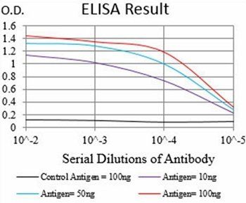
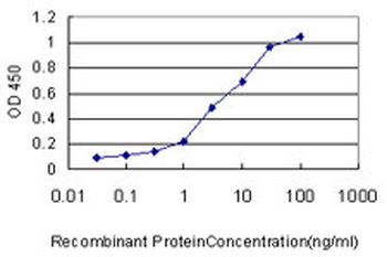
ELISA, IF, IHC-P, WB
Human
Mouse
Monoclonal
Unconjugated
100 μg - Item 1 of 6